
Page 41 - Dental Practice Vol 17 No.5_Neat
P. 41
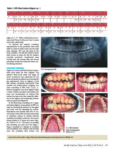
Table 1: IPR Chart before Aligner no. 1


             edges of 11, 21. While maintaining harmo-
             nious Soft Tissue Profile and Class I canine
             and molar relationship.
                To  alleviate  the  anterior  crowding,
             reproximation  of  the  posteriors  were  done
             distal to canine in both arches and the teeth
             were  retracted.  IPR  was  also  done  in  the
             lower anteriors. The bite was raised on lower
             third  molars  to  jump  the  bite  for  anterior
             crossbite  correction.  Attachments  were
             bonded  onto  the  canines,  first  and  second
             premolars in both the arches for better con-
             trol during retraction.

             TREATMENT PROGRESS
             Upper and lower polyvinyl siloxane impres-  FIG 2: Pre-treatment OPG
             sions  were  taken  for  clear  Aligners.  The
             patient’s  final  tooth  setup  and  stages  of
             tooth  movement  were  generated  by  the
             three-dimensional software and reviewed by
             the  orthodontist.  Prior  to  delivery  of  the
             first  aligner,  composite  attachments  were
             bonded  and  interproximal  stripping  was
             done  according  to  IPR  chart  (Table  1).
             Patient changed to new set of aligners every                          3a                               3b
             2 weeks. Patient was given instructions for
             wear  and  maintenance  of  Aligners.The
             patient was seen every four weeks for deliv-                                  3c                       3d
             ery of new aligners and monitoring of treat-
             ment progress and aligner fit.
                In the first phase, consisting of 14 upper
             and lower aligners, space gained by IPR was
             used for decrowding anteriors in the upper
             and  lower  arch  by  retracting  the  canines.
             Impressions were retaken after this. In sec-
             ond phase 0.2mm IPR was done only mesial
             to  maxillary  canines  to  further  alleviate            3e
             crowding of maxillary anteriors. The second
             phase consists of 12 upper and lower align-
             ers. (Figures 3a-3e). Correction of crossbite
             and  crowding  was  seen  post  the  Stage  2.
             Stage 3 consisted of 4 aligners in maxillary
             and mandibular arch. Buttons were bonded                      FIG 3: Mid treatment
             onto  the  maxillary  first  molars  and                      intraoral photographs
                                                                           (Aligner no. 9)

               Access this article online  https://dental-practice.biz/emagazine/dentalpractice/mobile/#p=40


                                                                         Dental Practice // May-June 2021 // Vol 17 No 5 41