Page 173 - vol21_editedversion2
P. 173
Inhibition zone chart for Bacillus cereus

                            7.0
                                     6.0 c       6.0 c       6.0 c        6.0 c
                            6.0
                          Inhibition zone (mm)   4.0                                   Bacillus cereus
                            5.0


                            3.0

                            2.0

                            1.0

                            0.0
                                    control     Formulation 1 Formulation 2 Formulation 3

                                                   Formulations

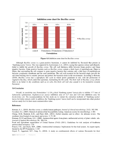
                                             Figure 4.6 Inhibition zone chart for Bacillus cereus

                  Although  Bacillus  cereus  is  a  gram-positive  bacterium,  it  cannot  be  inhibited  by  flavonoid  that  present  in
              ‘Sambung nyawa’ leaves. This was supported by Kaewseejan et al., (2012), saying that aqueous extract and ethanolic
              failed  to  inhibit  the  growth  of  Bacillus  cereus.  The  cell  wall  thickness  differ  between  Gram  positive  and  Gram
              negative bacteria. Gram positive bacteria such as  Bacillus cereus contain a thick layer of peptidoglycan; 20µm to
              80µm, that surrounding the cell compare to gram negative bacteria that contains only a thin layer of peptidoglycan
              between cytoplasmic membrane and the outer membrane. The cell wall essential for the bacterial shape, prevent the
              cell from bursting due to osmotic pressure and protect the bacterial strain in the environment. According to Bottone
              (2010),  Bacillus  cereus  has  a  saprophytic  life  cycle  in  which  spores  germinate  in  soil,  with  the  production  of
              vegetative Bacillus, which could then sporulate, maintaining the life cycle. The thick wall of Bacillus cereus allows
              them  to  do  better  in  dry  conditions  such  as on  soils, this  thick  cell  wall  also  caused  it  to  be  insensitive  towards
              flavonoid.

              5.0 Conclusion

                  Overall, in sensitivity test, Formulation 1 (1.50 g dried ‘Sambung nyawa’ leaves) able to inhibits 17.7 mm of
              Salmonella  typhimurium,  Staphylococcus  aureus  inhibition  zone  of  12.7  mm  and  12.0  mm  inhibition  zone  for
              Escherichia  coli.  The  recommendations  for this  study  are  to  variant  the  extraction  methods  of  ‘Sambung  nyawa’
              leaves for better extracts yield. In addition, the ‘Sambung nyawa’ leaves itself can be incorporated into other product
              such as candy for it to have more commercialise value.

              References

              Bottone, E. J., (2010). Bacillus cereus, a volatile human pathogen. Journal of clinical microbiology. 23(2). 382-398.
              Cavalieri, S.J., (2005). Manual of antimicrobial Susceptibility testing. American society for microbiology. 1-481
              Chirag,  M.M,  Shailesh,  K.M.,  and  Patel,  H.B.,  (2012).  Herbal  remedies  and  its  effect:  An  ultimate  review.  The
              southeast Asia Journal of case report and review. 3, 73- 88
              Dorman, H.J.D and Deans, S.G., (2000). Antimicrobial agents from plants: antibacterial activity of plant volatile  oils.
              Journal of Applied Microbiology, 88,  308-316.
              Food  and  Agriculture  organization  of  United  Nations  (FAO)  (2011).  Guidelines  for  risk  analysis  of  foodborne
              antimicrobial resistance. 1-29
              Francis, F.B., and Doyle, M.P., (2006). Antimicrobial resistance: Implications for the food system. An expert report,
              funded by the IFT Foundation. 1-185
              Goyal,  P.,  Aggarwal,  B.P.,  Garg.  S.,  (2010).  A  study  on  combinatorial  effects  of  various  flavonoids  for  their
              163 | O M I I C O T   –   V O L 2 1

