
Page 532 - Physics Coursebook 2015 (A level)
P. 532
bone
520
Cambridge International A Level Physics
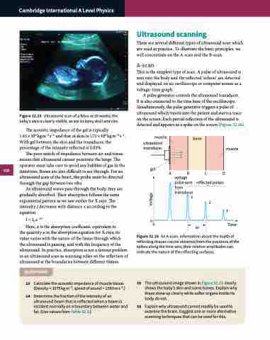
Figure 32.25 Ultrasound scan of a fetus at 20 weeks; the baby’s skin is clearly visible, as are its bony skull and ribs.
Ultrasound scanning
There are several different types of ultrasound scan which are used in practice. To illustrate the basic principles, we will concentrate on the A-scan and the B-scan.
A-scan
This is the simplest type of scan. A pulse of ultrasound is sent into the body and the reflected ‘echoes’ are detected and displayed on an oscilloscope or computer screen as a voltage–time graph.
A pulse generator controls the ultrasound transducer.
It is also connected to the time base of the oscilloscope. Simultaneously, the pulse generator triggers a pulse of ultrasound which travels into the patient and starts a trace on the screen. Each partial reflection of the ultrasound is detected and appears as a spike on the screen (Figure 32.26).
The acoustic impedance of the gel is typically 1.65×10⁶ kgm−2 s−1 and that of skin is 1.71×10⁶ kgm−2 s−1. With gel between the skin and the transducer, the percentage of the intensity reflected is 0.03%.
The poor match of impedance between air and tissue means that ultrasound cannot penetrate the lungs. The operator must take care to avoid any bubbles of gas in the intestines. Bones are also difficult to see through. For an ultrasound scan of the heart, the probe must be directed through the gap between two ribs.
As ultrasound waves pass through the body, they are gradually absorbed. Their absorption follows the same exponential pattern as we saw earlier for X-rays. The intensity I decreases with distance x according to the equation
I = I0 e−αx
Here, α is the absorption coefficient, equivalent to
the quantity μ in the absorption equation for X-rays; its value varies with the nature of the tissue through which the ultrasound is passing, and with the frequency of the ultrasound. In practice, absorption is not a serious problem in an ultrasound scan as scanning relies on the reflection of ultrasound at the boundaries between different tissues.
QUESTIONS
13 Calculate the acoustic impedance of muscle tissue. (Density = 1075 kg m−3, speed of sound = 1590 m s−1.)
14 Determine the fraction of the intensity of an ultrasound beam that is reflected when a beam is incident normally on a boundary between water and fat. (Use values from Table 32.3.)
muscle
ultrasound transducer
gel
00
muscle
ABCD
voltage pulse sent from transducer
reflected pulses
1
2
Δt
3 Time
Figure 32.26 An A-scan. Information about the depth of reflecting tissues can be obtained from the positions of the spikes along the time axis; their relative amplitudes can indicate the nature of the reflecting surfaces.
15 The ultrasound image shown in Figure 32.25 clearly shows the baby’s skin and some bones. Explain why these show up clearly while softer organs inside its body do not.
16 Explain why ultrasound cannot readily be used to examine the brain. Suggest one or more alternative scanning techniques that can be used for this.
Voltage
