
Page 533 - Physics Coursebook 2015 (A level)
P. 533
Chapter 32: Medical imaging
In Figure 32.26, pulses 1, 2 and 3 are reflected at
the various boundaries. Pulse 1 is the reflection at the muscle–bone boundary at B. Pulse 2 is the reflection at the bone–muscle boundary at C. The time Δt is the time taken for the ultrasound to travel twice the thickness of the bone. Finally, pulse 3 is the reflection at the muscle– air boundary at D. The thickness of the bone can be determined from this A-scan.
Each reflected pulse is analysed to determine the depth of the reflecting surface (from the time of echo) and the nature of the surface (from the amplitude of the reflected wave). A two-dimensional image is then built up on a screen by positioning dots to represent the position of the reflecting surfaces and with brightness determined by the intensity of the reflection, brighter dots indicating more reflected ultrasound (see Figure 32.27).
Figure 32.28 shows the result of a typical B-scan. Because it takes several seconds for the scanner to move across the body, problems can arise if the organs of interest are moving – this gives a blurred image.
time interval between pulses 1 and 2 = Δt
distance travelled by ultrasound
thickness of bone = 2
= cΔt 2
where c is the speed of the ultrasound in the bone (see Worked example 3).
Because ultrasound waves are gradually attenuated as they pass through the body (their energy is absorbed so that their amplitude and intensity decrease), the echoes from tissues deeper in the body are weaker and must be amplified.
A-scans are used for some straightforward procedures such as measuring the thickness of the eye lens.
WORKED EXAMPLE
3 In a particular A-scan, similar to Figure 32.26, the time interval between pulses 1 and 2 is 12 μs. The speed of ultrasound in bone is about 4000 m s−1. Determine the thickness of the bone.
Step1 Determinethedistancetravelledbythe ultrasound in the time interval of 12 μs.
distance = speed × time distance=4000×12×10−6 =4.8×10⁻²m
Step2 Calculatethethicknessofthebone.
Hint: The distance you have just calculated must be halved because the ultrasound has to travel through the bone twice.
thicknessofbone= 4.8×10⁻² 2
=2.4×10⁻²m(2.4cm)
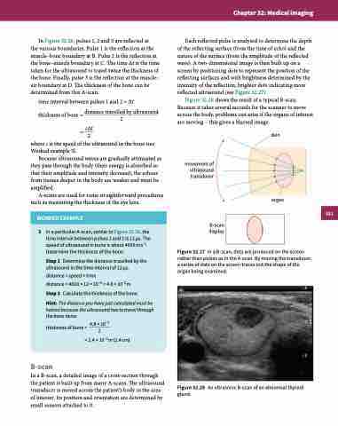
B-scan
In a B-scan, a detailed image of a cross-section through the patient is built up from many A-scans. The ultrasound transducer is moved across the patient’s body in the area of interest. Its position and orientation are determined by small sensors attached to it.
movement of ultrasound transducer
B-scan display
skin
organ
Figure 32.27 In a B-scan, dots are produced on the screen rather than pulses as in the A-scan. By moving the transducer, a series of dots on the screen traces out the shape of the organ being examined.
Figure 32.28 An ultrasonic B-scan of an abnormal thyroid gland.
521
