
Page 535 - Physics Coursebook 2015 (A level)
P. 535
Chapter 32: Medical imaging
A proton does not align itself directly along the external field. In practice, its magnetic axis rotates around the direction of the external field (Figure 32.31), just like the axis of a spinning top. This rotation or gyration action is known as precession.
resonance requires a system with a natural frequency of vibration; when it is stimulated with energy of the same frequency, it absorbs energy. In MRI, protons precessing about the strong external field are exposed to a burst or pulse of RF waves whose frequency equals the frequency of precession. Each proton absorbs a photon of RF energy and flips up into the higher energy state; this is nuclear magnetic resonance (Figure 32.32).
Now we come to the useful bit. The RF waves are switched off and the protons gradually relax into their lower energy state. As they do so, they release their excess energy in the form of RF waves. These can be detected, and the rate of relaxation tells us something about the environment of the protons.
In Figure 32.32, you can see that the relaxation of the protons follows an exponential decay pattern. Curves like this are characterised by two relaxation times:
■■ T1, the spin–lattice relaxation time, where the energy of the spinning nuclei is transferred to the surrounding ‘lattice’ of nearby atoms
■■ T2, the spin–spin relaxation time, where the energy is transferred to other spinning nuclei.
These relaxation times depend on the environment of the nuclei. For biological materials, it depends on their water content:
■■ Water and watery tissues (e.g. cerebrospinal fluid) have relaxation times of several seconds.
■■ Fatty tissues (e.g. white matter in the brain) have shorter relaxation times, several hundred milliseconds.
■■ Cancerous tissues have intermediate relaxation times.
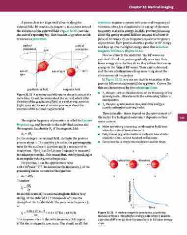
path of precession
axis of spin
gravitational field
path of spin precession
axis of spin spin
magnetic field
Figure 32.31 A spinning top (left) rotates about its axis; at the same time, its axis precesses about the vertical, which is the direction of the gravitational field. In a similar way, a proton (right) spins and its axis of rotation precesses about the direction of the external magnetic field.
The angular frequency of precession is called the Larmor frequency ω0, and depends on the individual nucleus and the magnetic flux density B0 of the magnetic field:
ω0 = γB0
So, the stronger the external field, the faster the protons precess about it. The quantity γ is called the gyromagnetic ratio for the nucleus in question and is a measure of its magnetism. (Note that the Larmor frequency is measured in radians per second. This means that, strictly speaking, it is an angular velocity, not a frequency.)
For protons, γ has the approximate value
2.68 × 10⁸ rad s−1 T−1. To determine the frequency f0 of the precessing nuclei, we can use the equation:
ω0 = 2πf0 Therefore:
f0 = λB0 2π
In an MRI scanner, the external magnetic field is very strong, of the order of 1.5 T (thousands of times the strength of the Earth’s field). The precession frequency f0 is:
f0 = 2.68×108 ×1.5 = 6.4×10⁷ Hz = 64MHz 2π
This frequency lies in the radio frequency (RF) region of the electromagnetic spectrum. You should recall that
absorption
relaxation
00 Time
Figure 32.32 In nuclear magnetic resonance, a spinning nucleus is flipped into a higher energy state when it absorbs a photon of RF energy; then it relaxes back to its lower energy state.
523
Energy of protons
