
Page 536 - Physics Coursebook 2015 (A level)
P. 536
524
Cambridge International A Level Physics
This means that different tissues can be distinguished by the different rates at which they release energy after they have been forced to resonate. That is the basis of medical applications of nuclear magnetic resonance.
QUESTIONS
19 Protons precess at a frequency of 42.6 MHz in an external field of magnetic flux density 1.0 T.
a Determine the frequency at which will they precess in a field of magnetic flux density 2.5 T.
b State the frequency of RF radiation that will cause the protons to resonate in this stronger magnetic field.
20 Figure 32.33 shows how the amplitude of RF waves coming from watery tissue varies after resonance. Copy the graph and add lines and labels to show the graphs you would expect to see for cancerous and fatty tissues.
watery tissue
00 Time
Figure 32.33 See Question 20.
MRI scanner
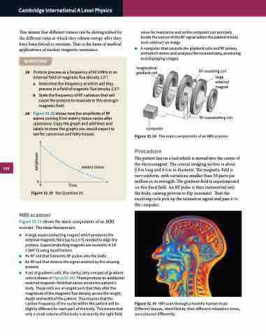
Figure 32.34 shows the main components of an MRI scanner. The main features are:
■■ A large superconducting magnet which produces the external magnetic field (up to 2.0 T) needed to align the protons. Superconducting magnets are cooled to 4.2 K (−269 °C) using liquid helium.
■■ An RF coil that transmits RF pulses into the body.
■■ An RF coil that detects the signal emitted by the relaxing
protons.
■■ A set of gradient coils. (For clarity, only one pair of gradient
value for resonance and so the computer can precisely locate the source of the RF signal within the patient’s body and construct an image.
■■ A computer that controls the gradient coils and RF pulses, and which stores and analyses the received data, producing and displaying images.
Amplitude
coils is shown in Figure 32.34.) These produce an additional external magnetic field that varies across the patient’s body. These coils are arranged such that they alter the magnitude of the magnetic flux density across the length, depth and width of the patient. This ensures that the Larmor frequency of the nuclei within the patient will be slightly different for each part of the body. This means that only a small volume of the body is at exactly the right field
Figure 32.35 MRI scan through a healthy human head. Different tissues, identified by their different relaxation times, are coloured differently.
longitudinal gradient coil
RF transmitting coil Figure 32.34 The main components of an MRI scanner.
Procedure
The patient lies on a bed which is moved into the centre of the electromagnet. The central imaging section is about 0.9 m long and 0.6 m in diameter. The magnetic field is very uniform, with variations smaller than 50 parts per million in its strength. The gradient field is superimposed on this fixed field. An RF pulse is then transmitted into the body, causing protons to flip (resonate). Then the receiving coils pick up the relaxation signal and pass it to the computer.
computer
RF receiving coil
large external magnet
