
Page 152 - ASOP ROT Study Guide
P. 152
www.orthopedicarizona.com
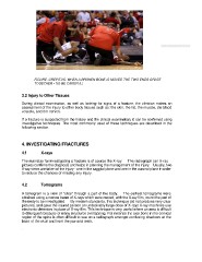
FIGURE. CREPITUS: WHEN A BROKEN BONE IS MOVED THE TWO ENDS GRATE
TOGETHER • SO BE CAREFUL!
3.2 Injury to Other Tissues
During clinical examination, as well as looking for signs of a fracture, the clinician makes an
assessment of the injury to other body tissues such as: the skin, the fat, the muscle, die blood
vessels, and the nerves.
If a fracture is suspected from the history and the clinical examination, it can be confirmed using
investigative techniques. The most commonly used of these techniques are described in the
following section.
4. INVESTIGATING FRACTURES
4.1 X-rays
The mainstay for investigating a fracture is of course the X-ray. The radiograph (an X-ray
picture) confirms the diagnosis and helps in planning the management of the injury. Usually, two
X-ray views are taken of the injury - one in the saggital plane and one in the coronal plane in order
to reduce the chances of missing any injury.
4.2 Tomograms
A tomogram is a view of "slice" through a part of the body. The earliest tomograms were
obtained using a narrow beam of X-rays which were moved, with the X-ray film, round the part of
the body to be investigated. By modern standards, this technique did not produce very clear
pictures, and gave the injured person an undesirably large dose of X-rays X-ray machines use
electronic detectors in place of X-ray film. This technique is very useful where an area is difficult
to distinguish because of many structures overlapping. For instance the axis bone in the cervical
region of the spine is often difficult to see on a radiograph amongst confusing shadows at the
base of the skull and from the jaw and teeth.
.