Page 332 - ECG dr shamol_Slide
P. 332
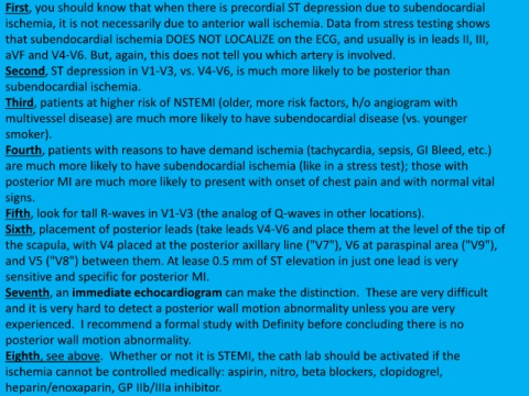
First, you should know that when there is precordial ST depression due to subendocardial
  ischemia, it is not necessarily due to anterior wall ischemia. Data from stress testing shows

  that subendocardial ischemia DOES NOT LOCALIZE on the ECG, and usually is in leads II, III,
  aVF and V4-V6. But, again, this does not tell you which artery is involved.

  Second, ST depression in V1-V3, vs. V4-V6, is much more likely to be posterior than

  subendocardial ischemia.
  Third, patients at higher risk of NSTEMI (older, more risk factors, h/o angiogram with

  multivessel disease) are much more likely to have subendocardial disease (vs. younger

  smoker).

  Fourth, patients with reasons to have demand ischemia (tachycardia, sepsis, GI Bleed, etc.)
  are much more likely to have subendocardial ischemia (like in a stress test); those with

  posterior MI are much more likely to present with onset of chest pain and with normal vital

  signs.
  Fifth, look for tall R-waves in V1-V3 (the analog of Q-waves in other locations).

  Sixth, placement of posterior leads (take leads V4-V6 and place them at the level of the tip of

  the scapula, with V4 placed at the posterior axillary line ("V7"), V6 at paraspinal area ("V9"),

  and V5 ("V8") between them. At lease 0.5 mm of ST elevation in just one lead is very
  sensitive and specific for posterior MI.

  Seventh, an immediate echocardiogram can make the distinction. These are very difficult

  and it is very hard to detect a posterior wall motion abnormality unless you are very
  experienced. I recommend a formal study with Definity before concluding there is no

  posterior wall motion abnormality.

  Eighth, see above. Whether or not it is STEMI, the cath lab should be activated if the
  ischemia cannot be controlled medically: aspirin, nitro, beta blockers, clopidogrel,

  heparin/enoxaparin, GP IIb/IIIa inhibitor.

