
Page 388 - ECG dr shamol_Slide
P. 388
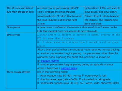
The SA node consists of             A central core of pacemaking cells (“P                  dysfunction  of This  cell leads to

         two main groups of cells:           cells”)  produce the sinus impulses                     sinus pauses and sinus arrest.

                                             transitional cells (“T cells”) that transmit  Failure of the T cells to transmit

                                             the sinus impulses out into the right                   the impulse. This leads to sino-

                                             atrium                                                  atrial exit block.
         Sinus pause                         A sinus pause is defined as the transient absence of sinus P waves on the

                                             ECG  that may last from two seconds to several minute

         Sinus arrest                        A sinus arrest is defined as absence of normal p-waves on the
                                             ECG for more than 2 seconds

                                             Or if sinus pause persist more than 2 second is called sinus

                                             arrest

                                             After a brief period either the sinoatrial node resumes normal pacing,
                                             or another pacemaker begins pacing. If a pacemaker other than the

                                             sinoatrial node is pacing the heart, this condition is known as

                                             an escape rhythm

                                             If no other pacemaker begins pacing during an episode of sinus

                                             arrest it becomes a cardiac arrest
         Three escape rhythm                 it is in the following order:

                                             1. Atrial escape (rate 60–80): normal P morphology is lost

                                             2. Junctional escape (rate 40–60): P is inverted or retrograde
                                             3. Ventricular escape (rate 20–40): no P wave, wide, abnormal QRS.