
Page 23 - ANZCP Gazette MAY 2014
P. 23
32oC, a target hematocrit on CPB of >0.22, mean arterial pressure (MAP) >50 mmHg and venous oxygen saturations maintained above 70%. The activated clotting time (ACT) was monitored during the bypass and maintained above 480 seconds. All patients received a general anaesthetic and �–stat acid/base management was used. The arterial filter purge line was connected via the sampling manifold to the venous inlet port and was open throughout CPB.
The venous line to cannula connection was de-aired or not, according to surgeon preference, prior to commencing CPB. During CPB, the oxygenator sweep gas composition and rate were titrated according to clinical needs. The HSVR was operated within the manufacturer’s recommended guidelines for minimum volume, with a low -volume alarm set at that threshold. Cardioplegia was delivered according to surgeon preferences using 4:1 blood cardioplegia. In accordance with standard practice, cardiotomy suction blood was sequestered for cell salvage at a later stage, delivered straight to the cell saver or added to the venous reservoir as required. Any blood vented from the left ventricle was returned to the venous reservoir or the cardiotomy reservoir, according to the perfusionist’s preference. Additional volume was administered as required via the dedicated ports on the venous reservoir or to the venous line, also according to the perfusionist’s preference. Drug additions were made via the sampling manifold to the HSVR in the perfusionist’s usual manner.
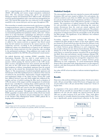
An emboli detection and counting (EDAC) quantifier (Luna Innovations, Roanoke, VA) was used to count emboli in the circuits. These devices utilize sonar-like technology to count and measure emboli passing the detector probes in the CPB circuit. Their accuracy and limitations have been assessed and described.6 Two sites on the CPB circuit were simultaneously monitored at fixed positions as follows: immediately upstream from the arterial filter and on the arterial line downstream from the arterial filter (Figure 1). The measurement of emboli immediately upstream and downstream of the filter facilitated precise evaluation of the removal of emboli by the arterial filter. Importantly, it largely mitigated the non-randomized design of the study because factors that could have differed between cases and which might have influenced the number and size of emboli reaching the filter (such as duration of CPB, the nature of the surgery and the management of cardiotomy blood) were controlled by counting those emboli upstream of the filter in all patients. Intraoperative emboli counting was started at the inception of CPB and ceased at the first termination of CPB. Data from both sensor sites were captured on the EDAC quantifier hard disk for each patient. Throughout the counting period, the EDAC quantifier recorded emboli counts within 10 μm size bands for both EDAC sites. Perfusion and procedural data were collected in the Stöckert Data Management System (DMS) patient file in keeping with normal practice.
Figure 1.
Statistical Analysis
The microemboli count data were reported as means with standard deviations (SD) and were treated as follows. For each patient, the emboli recorded at each of the two EDAC sites were totalled, both for each size band and for all sizes combined, and these counts were divided by the duration of the CPB period to produce a count per minute. The proportion of emboli removed by the arterial filter for each procedure was calculated by subtracting the count per minute downstream from the count per minute upstream and dividing the result by the count per minute upstream. The primary outcome measure was a comparison of the mean (for all procedures) proportion of emboli removed by the arterial filter in the 20 and 40 μm filter groups. The significance of any difference was evaluated with a two-tailed t test.
Secondary outcome measures included the following. We compared the filter groups in respect of the mean emboli counts per minute by 10 μm emboli size intervals (from 0 to 100 μm), both upstream and downstream of the filter. A two-tailed t test was used to evaluate between group differences in each comparison. We also sought evidence for any obvious haemodynamic disadvantage during use of the 20 micron filter by comparing the two groups with respect to arterial line pressure (measured 10 cm proximal to the filter), flow rates and systemic vascular resistance during CPB. These parameters were measured every 30 seconds and the pooled measurements for each group were averaged for comparison. Once again, a two-tailed t test was used to evaluate between group differences in each comparison. To avoid measurements during drops in flow (associated with clamp placement or at the surgeon’s request), only data from periods where flow was ≥1.2 L.min1.m−2 were analysed.
A p-value of 0.05 or less was taken to indicate statistical significance in all tests.
Results
Data were collected from 24 bypass procedures. Twelve procedures with a 40 μm arterial filter totaled 18.2 hours of bypass time and 12 procedures with a 20 μm arterial filter totaled 22.7 hours of bypass time.
A comparison of the mean emboli counts per minute upstream of both 20 μm and 40 μm arterial filters revealed no significant difference for any 10 μm emboli size interval between 10 and 100 μm (p-values between 0.23 and 0.54). Thus, both filters were exposed to virtually identical incoming emboli loads. There was a significantly greater proportional reduction in the mean
Arterial Filter
Circuit design used in this experiment, showing EDAC sensor positioning.
EDAC: emboli detection and classification quantifier; HSVR: hard-shell venous reservoir.
HSVR
EDACS Site 2
(Post Arteriak Filter)
Oxygenator
EDACS Site 1
(Pre Arteriak Filter)
MAY 2014 | www.anzcp.org
21
