
Page 69 - Human Umbilical Cord Mesenchymal Stem Cells
P. 69
340 DING ET AL.
To date, younger stem cells include fetal stem cells Cord Lining
obtained from amniotic fluid, the umbilical cord (UC), Umbilical cord lining cells have been isolated by the
and placenta (4,9,66). Fetal stem cells are mostly MSCs, explant method (25,56,57). Two kinds of cells, MSCs
and fetal MSCs from birth-associated tissues are gaining (CLMCs) and epithelial cells (CLECs), can be isolated from
popularity. MSCs derived from the umbilical cord can cord lining. The CLMCs are isolated from the subamnion
be obtained from the amniotic membrane, cord lining, region by dissecting out the Wharton’s jelly. Pieces of the
Wharton’s jelly, and perivascular region (Fig. 1). They outer envelope membranes are cultured with Connaught
are the focus of this review (Fig. 2). Medical Research Laboratories (CMRL) 1660 containing L-
glutamine and 10% fetal bovine serum (FBS) (33). Around
STEM CELLS DERIVED FROM DIFFERENT
20 million cells can be generated at passage 1 (38).
PARTS OF UMBILICAL CORD
The CLECs can be used for treating persistent corneal
The human umbilical cord starts developing on the
epithelial defects and as a skin cosmetic improvement,
fifth week of gestation and continues to grow until 50 cm
whereas CLMCs have been used for burn and diabetic
in length (75). Stem cells can be derived from various
ulcer wound healing (38). Since preclinical studies reveal
parts of the umbilical cord. All of these compartments
successful disease treatment, further exploration of the
have been described in the literature, including Wharton’s
utility of these cells is warranted.
jelly, cord lining, and the perivascular region.
BIOMARKER OF HUCMSCs
Wharton’s Jelly
Surface Markers
Most studies use UC MSCs from Wharton’s jelly
(2,9,74). Regarding isolation, there are two kinds of MSCs are positive for cluster of differentiation 29
methods: the explant method and the enzymatic digestion (CD29), CD44, CD90, CD73, CD105, and human leukocyte
method (50). In the explant method, the Wharton’s jelly antigen (HLA)-ABC. Conversely, MSCs are negative for the
3
is manually minced into 1–2-mm fragments after removal endothelial cell marker CD31; hematopoietic cell markers
of UC vessels. The fragments are undisturbed for 7 days CD34, CD45, and CD117; and HLA-DR (14,62). The sur-
to allow the stem cells to come out (9). However, the face markers of HUCMSCs are similar to those of MSCs
downside of the explant method is that the fragments (31), but they are negative for CD133 (31). Nevertheless,
often float in the medium. Moreover, this method may phenotypic characterization of HUCMSCs may be influ-
not provide a consistent number of MSCs. enced by the culture passage number, medium, and method.
In the enzymatic digestion method, the enzymes used
for digestion vary from collagenase to a combination of Embryonic Stem Cell Markers
collagenase and hyaluronidase with or without trypsin Octamer-binding transcription factor 4 (Oct4), Nanog,
(12,59,65). This method can provide more homogenous sex-determining region Y box 2 (Sox2), and Kruppel-like
cell populations and more consistent cell numbers com- factor 4 (KLF4) are expressed only at low levels in
pared to the explant method. HUCMSCs (27), suggesting that MSCs are primitive
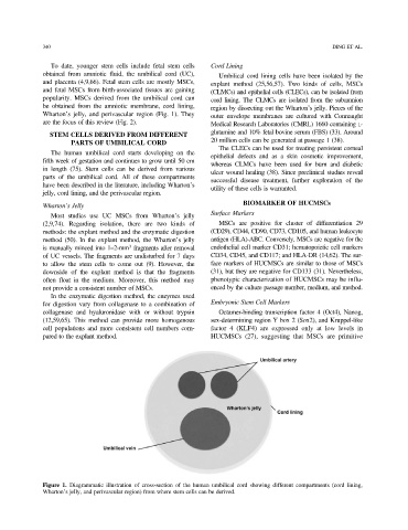
Figure 1. Diagrammatic illustration of cross-section of the human umbilical cord showing different compartments (cord lining,
Wharton’s jelly, and perivascular region) from where stem cells can be derived.