
Page 59 - Atlas of Small Animal CT and MRI
P. 59
Temporomandibular Joint 49
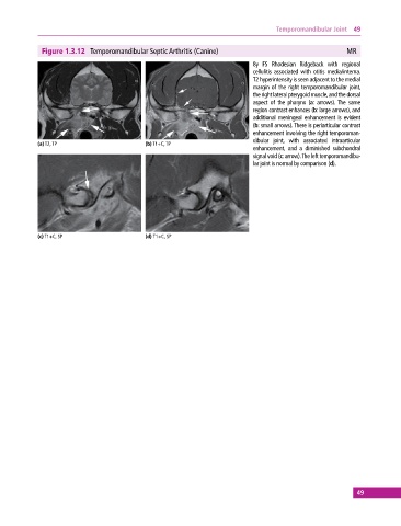
Figure 1.3.12 Temporomandibular Septic Arthritis (Canine) MR
8y FS Rhodesian Ridgeback with regional
cellulitis associated with otitis media/interna.
T2 hyperintensity is seen adjacent to the medial
margin of the right temporomandibular joint,
the right lateral pterygoid muscle, and the dorsal
aspect of the pharynx (a: arrows). The same
region contrast enhances (b: large arrows), and
additional meningeal enhancement is evident
(b: small arrows). There is periarticular contrast
enhancement involving the right temporoman-
dibular joint, with associated intraarticular
(a) T2, TP (b) T1+C, TP
enhancement, and a diminished subchondral
signal void (c: arrow). The left temporomandibu-
lar joint is normal by comparison (d).
(c) T1+C, SP (d) T1+C, SP
49