Page 99 - Atlas of Small Animal CT and MRI
P. 99

Globe  89


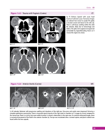
              Figure 1.6.3  Trauma with Proptosis (Canine)                                                CT
                                                                             3y FS Brittany Spaniel with acute head
                                                                             trauma of unknown cause. Transverse images
                                                                             are ordered from rostral to caudal. The globe
                                                                             is located cranial and lateral to the orbit (a).
                                                                             There is soft‐tissue swelling within the orbit
                                                                             (b: asterisk), likely due to hemorrhage and
                                                                             edema. The optic nerve appears enlarged and
                                                                             surrounded by hypoattenuating tissue as it
                                                                             crosses this region (b: arrow).


            (a) CT, TP                       (b) CT, TP


             (c) CT, DP                      (d) CT, DP


              Figure 1.6.4  Anterior Uveitis (Canine)                                                    MR


            (a) T1, DP                       (b) T2, DP                       (c) T1+C, DP
            1y M Labrador Retriever with progressive swelling and cloudiness of the right eye. Glaucoma and uveitis were diagnosed following a
            complete ophthalmic examination. There is increased signal intensity of the right anterior chamber on T1 images (a: arrow) compared to
            the normal eye. There is a similar but more subtle increase in vitreous attenuation in the same eye. On contrast‐enhanced images, there
            is marked enhancement of the fluid in the anterior chamber (c). The eye was enucleated after a corneal rupture, and plant material was
            found within the lens capsule.


                                                                                                             89
   94   95   96   97   98   99   100   101   102   103   104