
Page 407 - Atlas of Small Animal CT and MRI
P. 407
Thoracic wall and Diaphragm 397
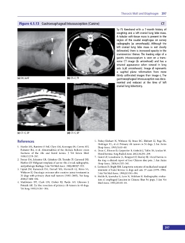
Figure 4.1.13 Gastroesophageal Intussusception (Canine) CT
5y FS Keeshond with a 7‐month history of
coughing and a left cranial lung lobe mass.
A tubular soft‐tissue mass is present in the
region of the caudal esophagus on survey
radiographs (a: arrowheads). Although the
left cranial lung lobe mass is not clearly
delineated, there is increased opacity in the
cranioventral thorax. The leading edge of a
gastric intussusceptum is seen on a trans-
verse CT image (b: arrowhead) and has a
striated appearance when viewed in long
axis (c,d: arrowheads). Image d represents
a sagittal plane reformation from more
thinly collimated images than image c. The
(a) DX, LLAT (b) CT+C, TP gastroesophageal intussusception was docu-
mented and reduced at the time of left
cranial lung lobectomy.
(c) CT+C, SP (d) CT+C, SP
References 5. Pirkey‐Ehrhart N, Withrow SJ, Straw RC, Ehrhart EJ, Page RL,
Hottinger HL, et al. Primary rib tumors in 54 dogs. J Am Anim
1. Hardie EM, Ramirez O 3rd, Clary EM, Kornegay JN, Correa MT, Hosp Assoc. 1995;31:65–69.
Feimster RA, et al. Abnormalities of the thoracic bellows: stress 6. Dean C, Etienne D, Carpentier B, Gielecki J, Tubbs RS, Loukas M.
fractures of the ribs and hiatal hernia. J Vet Intern Med. Hiatal hernias. Surg Radiol Anat. 2012;34:291–299.
1998;12:279–287. 7. Guiot LP, Lansdowne JL, Rouppert P, Stanley BJ. Hiatal hernia in
2. Feeney DA, Johnston GR, Grindem CB, Toombs JP, Caywood DD, the dog: a clinical report of four Chinese shar peis. J Am Anim
Hanlon GF. Malignant neoplasia of canine ribs: clinical, radiographic, Hosp Assoc. 2008;44:335–341.
and pathologic findings. J Am Vet Med Assoc. 1982;180:927–933. 8. Lorinson D, Bright RM. Long‐term outcome of medical and surgical
3. Liptak JM, Kamstock DA, Dernell WS, Monteith GJ, Rizzo SA, treatment of hiatal hernias in dogs and cats: 27 cases (1978–1996).
Withrow SJ. Oncologic outcome after curative‐intent treatment in J Am Vet Med Assoc. 1998;213:381–384.
39 dogs with primary chest wall tumors (1992–2005). Vet Surg. 9. Stickle R, Sparschu G, Love N, Walshaw R. Radiographic evalua-
2008;37:488–496. tion of esophageal function in Chinese Shar Pei pups. J Am Vet
4. Matthiesen DT, Clark GN, Orsher RJ, Pardo AO, Glennon J, Med Assoc. 1992;201:81–84.
Patnaik AK. En bloc resection of primary rib tumors in 40 dogs.
Vet Surg. 1992;21:201–204.
396 397