
Page 460 - Atlas of Small Animal CT and MRI
P. 460
450 Atlas of Small Animal CT and MRI
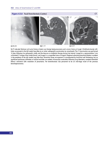
Figure 4.5.8 Focal Bronchiectasis (Canine) CT
(a) DX, DV (b) CT, TP (c) CT, TP
8y FS Labrador Retriever with prior history of plant awn foreign body pneumonia and a recent history of cough. Ill‐defined alveolar infil
trates are present in the left caudal lung lobe on an initial radiographic examination (a: arrowheads). The CT examination was performed
12 days following the radiographic study, and the dog was on antibiotic therapy during that interval. Image b is a representative 1 mm
collimated CT image of the caudal thorax, and image c is a magnified view of image b. Regional arborizing bronchial dilation is evident
in the periphery of the left caudal lung lobe (b,c). Bronchial lumen enlargement is accompanied by bronchial wall thickening, but no
significant pulmonary infiltrates or luminal exudates are evident. Microscopic evaluation following lung lobectomy revealed interstitial
fibrosis consistent with resolution of pneumonia. The bronchiectasis was presumed to be an end‐stage result of the previous
bronchopneumonia.
450