
Page 477 - Atlas of Small Animal CT and MRI
P. 477
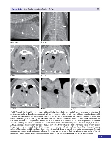
Figure 4.6.8  Left Cranial Lung Lobe Torsion (Feline)                                       CT


            (a) DX, RLAT                     (b) DX, DV


            (c) CT, TP                       (d) CT, TP


            (e) CT, TP                       (f) CT, TP                       (g) CT, TP
            13y MC Domestic Shorthair with 3‐month history of idiopathic chylothorax. Radiographic and CT images were acquired at the time of
            most recent evaluation (a–f) and 2 months previously (g). Images c–e were acquired through the cranial thorax and ordered from cranial
            to caudal. Image f is a magnified view of image e. Image g was acquired at approximately the same level as image e. Radiographs
            revealed air bronchograms and alveolograms (a,b: arrowheads) and a possible truncated left cranial lobe bronchus (a: arrow) character-
            istic of lung lobe torsion. CT images reveal a characteristic heterogeneous air and soft‐tissue lung pattern of pulmonary emphysema
            associated with lung lobe torsion (c,d: arrows). The origin of the left cranial lobar bronchus tapers and truncates abruptly (e,f: arrows).
            Although the images are windowed to best define the pulmonary parenchyma features and the lobar bronchus, pleural effusion can also
            be appreciated (c–e: arrowheads). On the CT image acquired 2 months previously (g), a moderate pleural effusion is present, causing
            collapse of the cranial and middle lung lobes. However, the left cranial lobe bronchus is clearly identified (g: arrow) and can be followed
            unimpeded peripherally on adjacent images, indicating the torsion was not present at that time. Microscopic examination of the left
            cranial lung following surgical excision revealed extensive coagulation necrosis and hemorrhage as a consequence of torsion.
                                                                                                             467