
Page 491 - Atlas of Small Animal CT and MRI
P. 491
Small Airways and Parenchyma 481
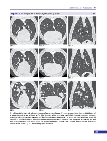
Figure 4.6.28 Progression of Pulmonary Metastasis (Canine) CT
(a) CT, TP (b) CT, TP (c) CT, DP
(d) CT, TP (e) CT, TP (f) CT, DP
(g) CT, TP (h) CT, TP (i) CT, DP
11y MC Labrador Retriever with pulmonary metastasis from an oral melanoma. CT images were acquired at the time of initial diagnosis
of primary disease (a–c) and at 73 days (d–f) and 157 days (g–i) following the initial scan. Multiple metastatic masses and nodules are
easily detected on representative transverse and dorsal plane images acquired at day 157 (g–i: arrowheads). By viewing comparable
images from the different dates in the vertical columns, one can appreciate the appearance and progression of metastasis over time.
Postmortem examination confirmed the presence of widespread pulmonary metastasis. The curvilinear densities near the diaphragm in
image c are due to diaphragmatic motion during image acquisition.
481