
Page 520 - Atlas of Small Animal CT and MRI
P. 520
510 Atlas of Small Animal CT and MRI
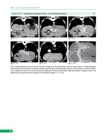
Figure 5.2.5 Congenital Intrahepatic Shunt—Left Divisional (Canine) CT
(a) CT+C, TP (b) CT+C, TP (c) CT+C, TP
(d) CT+C, TP (e) CT+C, TP (F) CT+C, DP
5mo FS Golden Retriever with poor growth. Transverse images (a–e) are ordered from caudal to cranial. There is a single intrahepatic
shunt arising from the portal vein at the porta hepatis and traveling to the left side (b–d: arrows). The vessel forms a curve that returns
to the caudal vena cava in the cranial liver near the diaphragm. The dorsal reformatted image demonstrates the typical shape of the
patent ductus venosus (f: arrows). See page 507 for Legend for Figures 5.2.1–5.2.18.
510