
Page 536 - Atlas of Small Animal CT and MRI
P. 536
526 Atlas of Small Animal CT and MRI
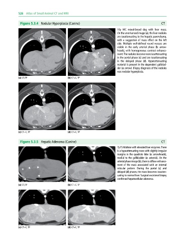
Figure 5.3.4 Nodular Hyperplasia (Canine) CT
10y MC mixed‐breed dog with liver mass.
On the unenhanced image (a), the liver nodules
are isoattenuating to the hepatic parenchyma,
with a suggestion of mass effect on the left
side. Multiple well‐defined round masses are
visible in the early arterial phase (b: arrow-
heads), with homogeneous contrast enhance-
ment. The nodules become more isoattenuating
in the portal phase (c) and are isoattenuating
in the delayed phase (d). Hyperattenuating
material is present in the dependent gallblad-
der (a: arrow). Biopsy diagnosis of the nodules
was nodular hyperplasia.
(a) CT, TP (b) CT+C, TP
(c) CT+C, TP (d) CT+C, TP
Figure 5.3.5 Hepatic Adenoma (Canine) CT
7y FS Maltese with elevated liver enzymes. There
is a hypoattenuating mass with slightly irregular
margins in the quadrate lobe (a: arrowheads),
medial to the gallbladder (a: asterisk). On the
arterial phase image (b), there is diffuse enhance-
ment of the mass associated with an internal
reticular pattern. During the portal (c) and
delayed (d) phases, the mass becomes isoatten-
uating to normal liver. Surgical excisional biopsy
confirmed hepatocellular adenoma.
(a) CT, TP (b) CT+C, TP
(c) CT+C, TP (d) CT+C, TP