
Page 543 - Atlas of Small Animal CT and MRI
P. 543
Hepatobiliary Disorders 533
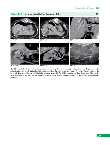
Figure 5.3.14 Lymphoma with Bile Duct Obstruction (Feline) CT
(a) CT+C, TP (b) CT+C, TP (c) CT+C, OP
(d) CT+C, OP (e) CT+C, DP (f) US, SP
12y MC Domestic Shorthair with hepatitis, anorexia, and vomiting. There is an irregular, isoattenuating mass (b,d,e: arrowheads)
obstructing the common bile duct and causing intrahepatic biliary distension (a,b,d: open arrows). The mass is located ventral to the
portal vein (b: arrow) and in close proximity to the duodenum (d: asterisk). The fluid‐attenuating, branching bile ducts are visible parallel
to the portal veins (c). The mass was also visible on ultrasound images near the duodenal papilla (f: calipers). Surgical biopsy confirmed
lymphoma.
533