
Page 82 - Feline Cardiology
P. 82
Chapter 9: Electrocardiography 79
A Diagnostic Testing
B
I II III aVR aVL aVF
I II III aVR aVL aVF
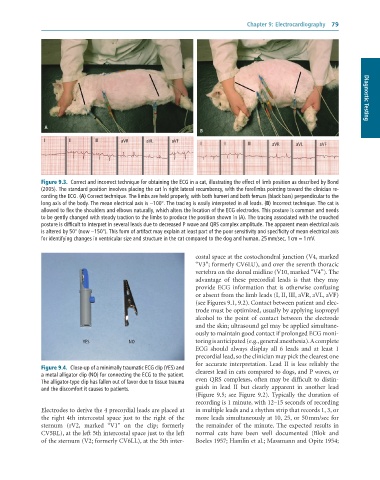
Figure 9.3. Correct and incorrect technique for obtaining the ECG in a cat, illustrating the effect of limb position as described by Bond
(2005). The standard position involves placing the cat in right lateral recumbency, with the forelimbs pointing toward the clinician re-
cording the ECG. (A) Correct technique. The limbs are held properly, with both humeri and both femurs (black bars) perpendicular to the
long axis of the body. The mean electrical axis is −100°. The tracing is easily interpreted in all leads. (B) Incorrect technique. The cat is
allowed to flex the shoulders and elbows naturally, which alters the location of the ECG electrodes. This posture is common and needs
to be gently changed with steady traction to the limbs to produce the position shown in (A). The tracing associated with the crouched
posture is difficult to interpret in several leads due to decreased P wave and QRS complex amplitude. The apparent mean electrical axis
is altered by 50° (now −150°). This form of artifact may explain at least part of the poor sensitivity and specificity of mean electrical axis
for identifying changes in ventricular size and structure in the cat compared to the dog and human. 25 mm/sec, 1 cm = 1 mV.
costal space at the costochondral junction (V4, marked
“V3”; formerly CV6LU), and over the seventh thoracic
vertebra on the dorsal midline (V10, marked “V4”). The
advantage of these precordial leads is that they may
provide ECG information that is otherwise confusing
or absent from the limb leads (I, II, III, aVR, aVL, aVF)
(see Figures 9.1, 9.2). Contact between patient and elec-
trode must be optimized, usually by applying isopropyl
alcohol to the point of contact between the electrode
and the skin; ultrasound gel may be applied simultane-
ously to maintain good contact if prolonged ECG moni-
YES NO toring is anticipated (e.g., general anesthesia). A complete
ECG should always display all 6 leads and at least 1
precordial lead, so the clinician may pick the clearest one
for accurate interpretation. Lead II is less reliably the
Figure 9.4. Close-up of a minimally traumatic ECG clip (YES) and clearest lead in cats compared to dogs, and P waves, or
a metal alligator clip (NO) for connecting the ECG to the patient.
The alligator-type clip has fallen out of favor due to tissue trauma even QRS complexes, often may be difficult to distin-
and the discomfort it causes to patients. guish in lead II but clearly apparent in another lead
(Figure 9.5; see Figure 9.2). Typically the duration of
recording is 1 minute, with 12–15 seconds of recording
Electrodes to derive the 4 precordial leads are placed at in multiple leads and a rhythm strip that records 1, 3, or
the right 4th intercostal space just to the right of the more leads simultaneously at 10, 25, or 50 mm/sec for
sternum (rV2, marked “V1” on the clip; formerly the remainder of the minute. The expected results in
CV5RL), at the left 5th intercostal space just to the left normal cats have been well documented (Blok and
of the sternum (V2; formerly CV6LL), at the 5th inter- Boeles 1957; Hamlin et al.; Massmann and Opitz 1954;