
Page 21 - Clinical Manual of Small Animal Endosurgery
P. 21
Rigid Endoscopy 9
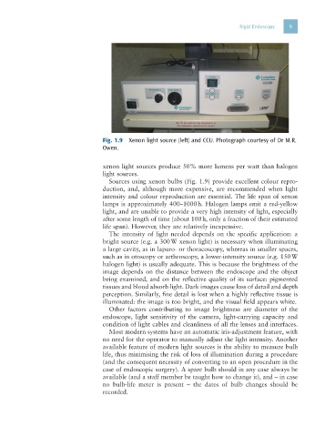
Fig. 1.9 Xenon light source (left) and CCU. Photograph courtesy of Dr M.R.
Owen.
xenon light sources produce 50% more lumens per watt than halogen
light sources.
Sources using xenon bulbs (Fig. 1.9) provide excellent colour repro-
duction, and, although more expensive, are recommended when light
intensity and colour reproduction are essential. The life span of xenon
lamps is approximately 400–1000 h. Halogen lamps emit a red-yellow
light, and are unable to provide a very high intensity of light, especially
after some length of time (about 100 h, only a fraction of their estimated
life span). However, they are relatively inexpensive.
The intensity of light needed depends on the specific application: a
bright source (e.g. a 300 W xenon light) is necessary when illuminating
a large cavity, as in laparo- or thoracoscopy, whereas in smaller spaces,
such as in otoscopy or arthroscopy, a lower-intensity source (e.g. 150 W
halogen light) is usually adequate. This is because the brightness of the
image depends on the distance between the endoscope and the object
being examined, and on the reflective quality of its surface: pigmented
tissues and blood absorb light. Dark images cause loss of detail and depth
perception. Similarly, fine detail is lost when a highly reflective tissue is
illuminated: the image is too bright, and the visual field appears white.
Other factors contributing to image brightness are diameter of the
endoscope, light sensitivity of the camera, light-carrying capacity and
condition of light cables and cleanliness of all the lenses and interfaces.
Most modern systems have an automatic iris-adjustment feature, with
no need for the operator to manually adjust the light intensity. Another
available feature of modern light sources is the ability to measure bulb
life, thus minimising the risk of loss of illumination during a procedure
(and the consequent necessity of converting to an open procedure in the
case of endoscopic surgery). A spare bulb should in any case always be
available (and a staff member be taught how to change it), and – in case
no bulb-life meter is present – the dates of bulb changes should be
recorded.