
Page 294 - Clinical Manual of Small Animal Endosurgery
P. 294
282 Clinical Manual of Small Animal Endosurgery
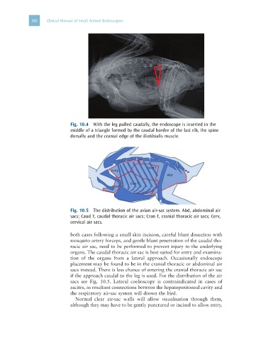
Fig. 10.4 With the leg pulled caudally, the endoscope is inserted in the
middle of a triangle formed by the caudal border of the last rib, the spine
dorsally and the cranial edge of the iliotibialis muscle.
Fig. 10.5 The distribution of the avian air-sac system. Abd, abdominal air
sacs; Caud T, caudal thoracic air sacs; Cran T, cranial thoracic air sacs; Cerv,
cervical air sacs.
both cases following a small skin incision, careful blunt dissection with
mosquito artery forceps, and gentle blunt penetration of the caudal tho-
racic air sac, need to be performed to prevent injury to the underlying
organs. The caudal thoracic air sac is best suited for entry and examina-
tion of the organs from a lateral approach. Occasionally endoscope
placement may be found to be in the cranial thoracic or abdominal air
sacs instead. There is less chance of entering the cranial thoracic air sac
if the approach caudal to the leg is used. For the distribution of the air
sacs see Fig. 10.5. Lateral coelioscopy is contraindicated in cases of
ascites, as resultant connections between the hepatoperitoneal cavity and
the respiratory air-sac system will drown the bird.
Normal clear air-sac walls will allow visualisation through them,
although they may have to be gently punctured or incised to allow entry.