
Page 93 - Clinical Manual of Small Animal Endosurgery
P. 93
Operative Arthroscopy 81
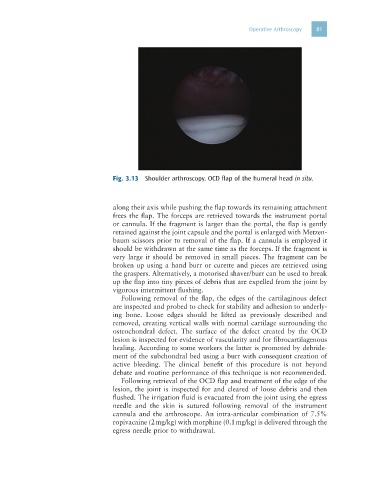
Fig. 3.13 Shoulder arthroscopy. OCD flap of the humeral head in situ.
along their axis while pushing the flap towards its remaining attachment
frees the flap. The forceps are retrieved towards the instrument portal
or cannula. If the fragment is larger than the portal, the flap is gently
retained against the joint capsule and the portal is enlarged with Metzen-
baum scissors prior to removal of the flap. If a cannula is employed it
should be withdrawn at the same time as the forceps. If the fragment is
very large it should be removed in small pieces. The fragment can be
broken up using a hand burr or curette and pieces are retrieved using
the graspers. Alternatively, a motorised shaver/burr can be used to break
up the flap into tiny pieces of debris that are expelled from the joint by
vigorous intermittent flushing.
Following removal of the flap, the edges of the cartilaginous defect
are inspected and probed to check for stability and adhesion to underly-
ing bone. Loose edges should be lifted as previously described and
removed, creating vertical walls with normal cartilage surrounding the
osteochondral defect. The surface of the defect created by the OCD
lesion is inspected for evidence of vascularity and for fibrocartilagenous
healing. According to some workers the latter is promoted by debride-
ment of the subchondral bed using a burr with consequent creation of
active bleeding. The clinical benefit of this procedure is not beyond
debate and routine performance of this technique is not recommended.
Following retrieval of the OCD flap and treatment of the edge of the
lesion, the joint is inspected for and cleared of loose debris and then
flushed. The irrigation fluid is evacuated from the joint using the egress
needle and the skin is sutured following removal of the instrument
cannula and the arthroscope. An intra-articular combination of 7.5%
ropivacaine (2 mg/kg) with morphine (0.1 mg/kg) is delivered through the
egress needle prior to withdrawal.