
Page 133 - Differential Diagnosis in Small Animal Cytology, The Skin and Subcutis
P. 133
er 8
Chapt
120
VetBooks.ir
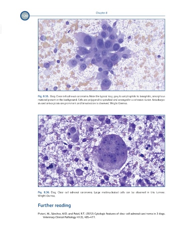
Fig. 8.35. Dog. Clear cell adnexal carcinoma. Note the typical lacy, grey to amphophilic to basophilic, amorphous
material present in the background. Cells are polygonal to spindloid and arranged in a cohesive cluster. Anisokaryo-
sis and anisocytosis are prominent and binucleation is observed. Wright-Giemsa.
Fig. 8.36. Dog. Clear cell adnexal carcinoma. Large multinucleated cells can be observed in this tumour.
Wright-Giemsa.
Further reading
Piviani, M., Sánchez, M.D. and Patel, R.T. (2012) Cytologic features of clear cell adnexal carcinoma in 3 dogs.
Veterinary Clinical Pathology 41(3), 405–411.