
Page 42 - Differential Diagnosis in Small Animal Cytology, The Skin and Subcutis
P. 42
Skin and Subcutis Anatomy
29
• Sac wall, apocrine and sebaceous cells in cats. Ducts and sacs are lined by stratified
squamous epithelium.
VetBooks.ir • Perianal gland epithelial cells (also called hepatoid glands) are modified sebaceous
epithelial cells. They are mostly found around the anus but also near the prepuce, on
the tail, flank and groin.
• Hair follicles
Hair follicles consist of an invagination of the epidermis into the dermis. They are respon-
sible for the formation of the hair, which is a modified keratinized structure. They are com-
posed of the following parts:
• Infundibulum: superficial portion, from the epidermis to the opening of the sebaceous duct.
• Isthmus: intermediate portion, from the opening of the sebaceous duct to the insertion
of the arrector pili muscle.
• Inferior segment: deep portion from the insertion of the arrector pili muscle to the base
of the follicle.
5.2 Main Cell Types Observed on Cytology
Different structures and types of epithelial and mesenchymal cells can be seen upon aspiration
of cutaneous and subcutaneous lesions.
Epithelial elements
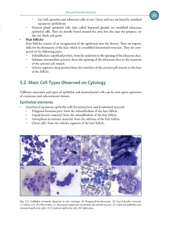
• Anucleated squamous epithelial cells (keratinocytes) and keratinized material:
• Polygonal keratinocytes: from the infundibulum of the hair follicle.
• Liquid keratin material: from the infundibulum of the hair follicle.
• Amorphous keratinous material: from the isthmus of the hair follicle.
• Ghost cells: from the inferior segment of the hair follicle.
(A) (B) (C) (D)
(E) (F) (G) (H)
Fig. 5.3. Epithelial elements observed in skin cytology. (A) Polygonal keratinocytes. (B) Liquid keratin material.
(C) Ghost cells. (D) Hair shafts. (E) Nucleated squamous epithelial cells (keratinocytes). (F) Cuboidal epithelial cells
(basaloid epithelial cells). (G) Columnar epithelial cells. (H) Sebocytes.