
Page 88 - Differential Diagnosis in Small Animal Cytology, The Skin and Subcutis
P. 88
Cysts, Tumour-like Lesions and Response to Tissue Injury
75
Pearls and Pitfalls
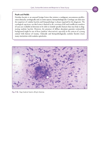
VetBooks.ir Nodular fasciitis is an unusual benign lesion that mimics a malignant sarcomatous prolifer-
ation clinically, cytologically and, in some aspects, histopathologically. Cytology can only raise
the suspicion of nodular fasciitis and histopathology is always required for the diagnosis. The
cytological experience on this lesion is limited in the veterinary field and insufficient numbers
of cases are available in literature to be able to identify specific features that may help in diag-
nosing nodular fasciitis. However, the presence of diffuse abundant granular eosinophilic
background might be one of these (authors’ observation), especially in the context of a young
animal with history of trauma. Clinically and histopathologically, nodular fasciitis shares
many similarities with nodular episcleritis.
Fig. 7.16. Dog. Nodular fasciitis. Wright-Giemsa.