
Page 53 - Rapid Review of ECG Interpretation in Small Animal Practice, 2nd Edition
P. 53
Holter Monitoring
VetBooks.ir
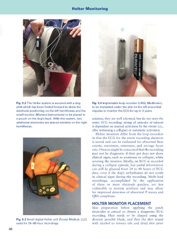
Fig. 5.2 The Holter system is secured with a dog Fig. 5.4 Implantable loop recorder (LINQ, Medtronic),
shirt which has been folded forward to show the to be implanted under the skin on the left precordial
electrode positioning on the left hemithorax and the impulse to monitor the ECG for up to 3 years.
small monitor (Mortara Instruments) to be placed in
a pouch on the dog’s back. With this system, two sedation, they are well tolerated, but do not store the
additional electrodes are placed similarly on the right entire ECG recording; saving of episodes of interest
hemithorax. is dependent on manual activation by the owner (i.e.,
after witnessing a collapse) or automatic activation.
Holter monitors differ from the loop recorders
in that the ECG for the entire recording duration
is stored and can be evaluated for abnormal beat
counts, maximum, minimum, and average heart
rate. Owners might be concerned that the recording
may not be diagnostic if their pet does not show
clinical signs, such as weakness or collapse, while
wearing the monitor. Ideally, an ECG is recorded
during a collapse episode, but useful information
can still be gleaned from 24 to 48 hours of ECG
data, even if the dog’s arrhythmias do not result
in clinical signs during the recording. Multi-lead
recordings, accomplished by the application
of three or more electrode patches, are less
vulnerable to motion artefacts and may allow
for improved detection of abnormal P waves and
QRS complexes.
HOLTER MONITOR PLACEMENT
Skin preparation before applying the patch
electrodes is critical to obtain a diagnostic ECG
recording. Hair needs to be clipped using the
Fig. 5.3 Small digital Holter unit (Forest Medical, LLC) shortest possible blade, and then the skin wiped
used for 24–48-hour recordings. with alcohol to remove oils and dried skin prior
40