Page 266 - Fluid, Electrolyte, and Acid-Base Disorders in Small Animal Practice
P. 266
Metabolic Acid-Base Disorders   257


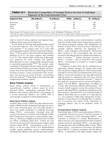
              TABLE 10-1       Electrolyte Composition of Luminal Fluid at the End of Individual
                               Segments of the Gastrointestinal Tract
            Segment End            Na (mEq/L)          K (mEq/L)          HCO 3 (mEq/L)           Cl (mEq/L)


            Duodenum                     60                 15                   15                    60
            Jejunum                     140                  6                   30                   100
            Ileum                       140                  8                   70                    60
            Colon                        40                 90                   30                   15*

            From Sleisinger MH, Fordtran JS, editors. Gastrointestinal diseases, 3rd ed. Philadelphia: WB Saunders, 1983: 258.
            *The large anion gap in luminal fluid at the end of the colon is caused by the presence of organic anions resulting from bacterial metabolism. These

            organic anions represent functional base loss in the stool because they could have been metabolized in the body to yield HCO 3 .
            rather is mixed if volume depletion and impaired tissue  citrate concentration), bone demineralization (resulting
            perfusion lead to lactic acid accumulation.         from loss of bone buffer stores during chronic acidosis),
              In one study of 134 dogs with gastroenteritis caused  and urinary potassium wasting with hypokalemia are
                                                                features of distal RTA in human patients. Mutations in
            by parvoviral infection, only 13% had low total CO 2

            concentrations. 121  In another study of 17 dogs with  cytosolic carbonic anhydrase, the basolateral Cl /

                                                                                                    þ
            parvoviral gastroenteritis, 59% had normal pH at presen-  HCO 3 anion exchanger, and luminal H -ATPase that
            tation. 108  In the animals with abnormal blood gas results,  affect function of the a-intercalated cells have been
            alkalemia (6 of 17) was more common than acidemia   associated with inherited forms of distal renal tubular aci-
                                                                                173
            (1 of 17). The majority (64%) of the dogs in this study  dosis in humans.  Urinary fractional excretion of
            were presented for both vomiting and diarrhea.      HCO 3    is normal (<5%) in distal RTA when plasma
            Hypochloremia is more common than hyperchloremia    HCO 3    concentration is increased to normal by alkali
            in parvoviral gastroenteritis. 108,121  In another study  administration.
            consisting of 25 puppies with parvoviral enteritis, plasma  A diagnosis of distal RTA may be confirmed by an
            concentrations of sodium, potassium, chloride, and bicar-  ammonium chloride tolerance test during which urine
            bonate were lower than those of control dogs; however,  pH is monitored (using a pH meter) before and at hourly
            increases in serum L-lactate concentration were uncom-  intervals for 5 hours after oral administration of 0.2 g/kg
            mon, and increases in serum D-lactate concentration  NH 4 Cl. Under such conditions, the urine pH of normal
            were not observed. 169  Most dogs in this study had mild  dogs decreased to a minimum value of 5.16 at 4 hours
            compensated metabolic acidosis.                     after administration of ammonium chloride. 214  Dogs in
                                                                this study also developed systemic acidosis (pH approxi-
            Renal Tubular Acidosis
                                                                mately 7.22 and HCO 3  approximately 14 mEq/L at
            Renal tubular acidosis (RTA) is characterized by    2 hours after ammonium chloride administration). The
            hyperchloremic metabolic acidosis caused by either  amount of alkali required to correct the acidosis in human

            decreased HCO 3   reabsorption (proximal RTA) or    patients with distal RTA is variable but typically less than
            defective acid excretion (distal RTA) in the presence of  that required in proximal RTA. The required dosage of
            a normal glomerular filtration rate (GFR). RTA is   alkali in distal RTA may be as little as 1 mEq/kg/day
            uncommonly recognized in small animal practice.     (i.e., that required to offset daily endogenous acid pro-
                                                                duction) or more than 2 to 4 mEq/kg/day. A combina-
            Distal Renal Tubular Acidosis                       tion of potassium and sodium citrate (depending on
            In distal (classic or type 1) RTA, the urine cannot be max-  potassium balance) may be the preferred source of
                                                                     196
            imally acidified because of impaired hydrogen ion secre-  alkali.
            tion in the collecting ducts, and urine pH typically is
            above 6.0, despite moderately to markedly decreased  Proximal Renal Tubular Acidosis

            plasma HCO 3   concentration. Increased urine pH    In proximal (type 2) RTA, renal reabsorption of HCO 3
            (>6.0) in the presence of acidosis is the hallmark of distal  is markedly reduced and urinary fractional excretion of

            RTA. Urinary tract infection by a urease-positive organ-  HCO 3 is increased (>15%) when plasma HCO 3 con-

            ism (e.g., Proteus sp., Staphylococcus aureus) must be ruled  centration is increased to normal. Bicarbonaturia is
            out before considering distal RTA. Urinary net acid  absent and urine pH is appropriately low when metabolic
            excretion is decreased, but bicarbonaturia usually is mild  acidosis is present and plasma HCO 3 concentration is


            because urinary HCO 3  concentration is only 1 to 3  decreased because distal acidifying ability is intact. When
            mEq/L in the pH range of 6.0 to 6.5. Nephrolithiasis  plasma HCO 3 concentration is decreased, the filtered

            (usually calcium phosphate stones), nephrocalcinosis  load of HCO 3 is reduced, and almost all of the filtered


            (resulting from alkaline urine pH and decreased urinary  HCO 3 is reabsorbed in the distal tubules, despite the

