
Page 625 - The Toxicology of Fishes
P. 625
Toxicity Resistance 605
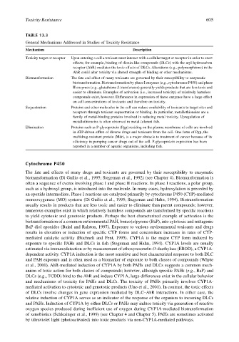
TABLE 13.3
General Mechanisms Addressed in Studies of Toxicity Resistance
Mechanism Description
Toxicity target or receptor Upon entering a cell a toxicant must interact with a cellular target or receptor in order to exert
effects; for example, binding of dioxin-like compounds (DLCs) with the aryl hydrocarbon
receptor (AhR) mediates the toxic effects of DLCs. Alterations (e.g., polymorphisms) in the
AhR could alter toxicity via altered strength of binding or other mechanisms.
Biotransformation The fate and effect of many toxicants are governed by their susceptibility to enzymatic
biotransformation. Biotransformation by phase I enzymes (e.g., cytochromes P450) and phase
II enzymes (e.g., glutathione S-transferases) generally yields products that are less toxic and
easier to eliminate. Examples of activation (i.e., increased toxicity) of relatively harmless
compounds exist, however. Differences in expression of these enzymes have a large effect
on cell concentrations of toxicants and therefore on toxicity.
Sequestration Proteins and other molecules in the cell can reduce availability of toxicants to target sites and
receptors through toxicant sequestration or binding. In particular, metallothioneins are a
family of metal-binding proteins involved in reducing metal toxicity. Upregulation of
metallothioneins is often observed in metal-tolerant fish.
Elimination Proteins such as P-glycoprotein (Pgp) residing on the plasma membrane of cells are involved
in ATP-driven efflux of diverse drugs and toxicants from the cell. One form of Pgp, the
multidrug-resistant protein (Mdr), is a major obstacle to treatment of cancer because of its
efficiency in pumping cancer drugs out of the cell. P-glycoprotein expression has been
reported in a number of aquatic organisms, including fish.
Cytochrome P450
The fate and effects of many drugs and toxicants are governed by their susceptibility to enzymatic
biotransformation (Di Guilio et al., 1995; Stegeman et al., 1992) (see Chapter 4). Biotransformation is
often a sequence of events involving phase I and phase II reactions. In phase I reactions, a polar group,
such as a hydroxyl group, is introduced into the molecule. In many cases, hydroxylation is preceded by
an epoxide intermediate. Phase I reactions are catalyzed primarily by cytochrome P450 (CYP)-mediated
monooxygenase (MO) systems (Di Guilio et al., 1995; Stegeman and Hahn, 1994). Biotransformation
usually results in products that are less toxic and easier to eliminate than parent compounds; however,
numerous examples exist in which relatively harmless compounds are transformed by specific reactions
to yield cytotoxic and genotoxic products. Perhaps the best characterized example of activation is the
biotransformation of a common environmental PAH, benzo(a)pyrene (BaP), into cytotoxic and mutagenic
BaP diol epoxides (Baird and Ralston, 1997). Exposure to various environmental toxicants and drugs
results in elevation or induction of specific CYP forms and concomitant increases in rates of CYP-
mediated catalytic activity (Buchneli and Fent, 1995). CYP1A is the major CYP form induced by
exposure to specific PAHs and DLCs in fish (Stegeman and Hahn, 1994). CYP1A levels are usually
estimated via immunodetection or by measurement of ethoxyresorufin-O-deethylase (EROD), a CYP1A-
dependent activity. CYP1A induction is the most sensitive and best characterized response to both DLC
and PAH exposure and is often used as a biomarker of exposure to both classes of compounds (Whyte
et al., 2000). AhR-mediated induction of CYP1A by both PAHs and DLCs suggests a common mech-
anism of toxic action for both classes of compounds; however, although specific PAHs (e.g., BaP) and
DLCs (e.g., TCDD) bind to the AhR and induce CYP1A, large differences exist in the cellular behavior
and mechanisms of toxicity for PAHs and DLCs. The toxicity of PAHs primarily involves CYP1A-
mediated activation to cytotoxic and genotoxic products (Uno et al., 2001). In contrast, the toxic effects
of DLCs involve changes in gene expression mediated by DLC–AhR interactions. In either case, the
relative induction of CYP1A serves as an indicator of the response of the organism to incoming DLCs
and PAHs. Induction of CYP1A by either DLCs or PAHs may induce toxicity via generation of reactive
oxygen species produced during inefficient use of oxygen during CYP1A-mediated biotransformation
of xenobiotics (Schlezinger et al., 1999) (see Chapter 4 and Chapter 5). PAHs are sometimes activated
by ultraviolet light (photoactivated) into toxic products via non-CYP1A-mediated pathways.