
Page 713 - The Toxicology of Fishes
P. 713
Biomarkers 693
MDR
OH
OH OH
SG
Phase II O
GST
OH
OH
Phase I O
CYP1A
DNA Adduct
Damage
OH HSP
Proteolysis
S-Protein
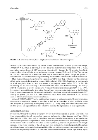
FIGURE 16.3 Relationship between phase I and phase II biotransformation and cellular responses.
aromatic hydrocarbons but induced by various cellular and xenobiotic oxidants (Leaver and George,
1998; Scott et al., 1992). In this way, it is quite likely that planar aromatic compounds, such as PCBs,
may induce certain forms of GST expression bifunctionally—through the Ah receptor (xenobiotic
response element) or oxidative stress (antioxidant response element) (Forlin et al., 1996). Thus, the use
of GST as a biomarker of exposure or effect may be limited unless specific assays and probes of
well-characterized isoforms are used together to help understand the relevance of induction or repression.
Many studies in humans have shown that expression of GSTs from the µ subfamily may have dramatic
effects on the susceptibility to various cancers (Seidegard et al., 1986, l990). In addition to proteins such
as MDR, neoplastic tissue has also been shown to overexpress various µ-class GSTs. In fish, this
relationship has not been as strong as that observed with MDR. Few studies examining GST activity
(CDNB conjugation) in hepatic lesions have documented consistent relationships (Kirby et al., 1990).
In a study of resistant Fundulus heteroclitus from a highly creosote-contaminated area in the Elizabeth
River, it was demonstrated that this population of fish had significant (sixfold) elevations in hepatic GST
activity and protein (Van Veld et al., 1991); however, unlike MDR levels, expression of GST did not
seem to differ between neoplastic or normal tissue.
Although UDPGT and GST may be used in certain instances to verify exposure to Ah receptor agonists,
their use as biomarkers of exposure is secondary to their use as biomarkers of effect (oxidative stress)
and susceptibility (particularly homologous µ-class GSTs). Clearly, many more characterization studies
in other species are required before these latter two uses may be implemented in field experiments.
Antioxidant Enzymes
Because oxidative insult can be an endogenous process that occurs normally in specific areas of the cell
(i.e., mitochondria), the cell has evolved numerous defenses to oxidant damage (see Figure 16.2).
Quantitatively, cellular thiols such as glutathione serve an extremely important role in maintaining the
cellular redox potential during oxidative stress. Several enzymes are involved in maintaining glutathione
in the reduced state. Glutathione reductase and the synthesizing enzymes of glutathione are strongly
regulated by the redox potential of the cell and have been postulated to be used as potential biomarkers
of oxidant damage and potentially of susceptibility. Channel catfish, for example, are significantly more
resistant to the pathological effects of oxidative stress than bullhead and contain significantly higher