Page 178 - Feline Cardiology
P. 178
178  Section D: Cardiomyopathies


              Pathophysiology                                    SIGNALMENT
              A stiffened left ventricle leads to elevated left ventricular   This is an adult onset disease. The average age at diag-
              diastolic pressure, dilation of the left atrium, pulmonary   nosis  has  been  reported  as  7 ± 3  years  (Ferasin  et  al.
              venous  congestion,  and  left  heart  failure.  Some  cats   2003).  Specific  breed  predispositions  have  not  been
              appear to develop pulmonary hypertension, and severe   identified,  although  it  has  been  reported  in  Birmans,
              right atrial dilation may be noted. Many cats have greatly   Siamese, and Persians as well as domestic shorthair and
              dilated left and right atria and develop thromboembolic   longhair (Meurs et al. 2000; Ferasin et al. 2003).
              episodes  as  well  as  atrial  tachyarrhythmias  including
              atrial fibrillation (Côté et al. 2004).            HISTORY AND CHIEF COMPLAINT
      Cardiomyopathies  Heart weight and heart weight to body weight are gener-  dyspnea, tachypnea, and anorexia. Restrictive cardiomy-
              Pathology
                                                                 The  presenting  complaints  may  be  vague  and  include
                                                                 opathy does have a high incidence of secondary throm-
              ally mildly to moderately increased (Fox 2004). The left
                                                                 boembolic disease, so hindlimb paralysis may be noted.
              ventricular wall thickness should be normal, but there
              may be some regional areas of thinning or hypertrophy.
              The left atrium is typically severely dilated. The left ven-
              tricular endocardium may be covered with an opaque,   PHYSICAL EXAMINATION
                                                                 A gallop sound has been suggested as a common auscul-
              white-gray fibrous material, particularly in the left ven-  tatory abnormality in some reports although a soft heart
              tricular outflow or on the papillary muscles. Some cats   murmur  is  often  heard  over  the  left  apex  or  at  the
              will  have  an  irregular  endocardial  fibrosis  in  the  left   sternum as well. In one report, a murmur was recorded
              ventricle  that  bridges  to  the  interventricular  septum   in 36% of the cases (Ferasin et al. 2003). A tachyarrhyth-
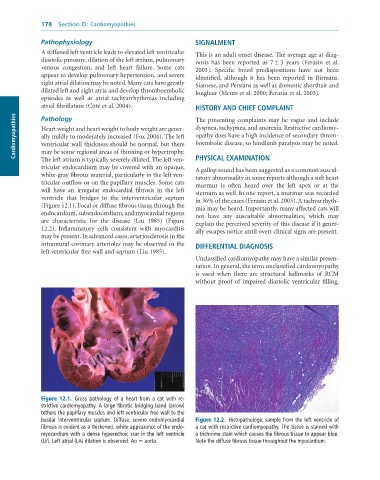
              (Figure 12.1). Focal or diffuse fibrous tissue through the   mia may be heard. Importantly, many affected cats will
              endocardium, subendocardium, and myocardial regions   not  have  any  auscultable  abnormalities,  which  may
              are  characteristic  for  the  disease  (Liu  1985)  (Figure   explain the perceived severity of this disease if it gener-
              12.2).  Inflammatory  cells  consistent  with  myocarditis   ally escapes notice until overt clinical signs are present.
              may be present. In advanced cases, arteriosclerosis in the
              intramural coronary arterioles may be observed in the   DIFFERENTIAL DIAGNOSIS
              left ventricular free wall and septum (Liu 1985).
                                                                 Unclassified cardiomyopathy may have a similar presen-
                                                                 tation. In general, the term unclassified cardiomyopathy
                                                                 is  used  when  there  are  structural  hallmarks  of  RCM
                                                                 without proof of impaired diastolic ventricular filling.

                                Ao

                                              LA


                                    LV


              Figure  12.1.  Gross	 pathology	 of	 a	 heart	 from	 a	 cat	 with	 re-
              strictive	cardiomyopathy.	A	large	fibrotic	bridging	band	(arrow)
              tethers	the	papillary	muscles	and	left	ventricular	free	wall	to	the
              basilar	interventricular	septum.	Diffuse,	severe	endomyocardial	  Figure 12.2.  Histopathologic	sample	from	the	left	ventricle	of
              fibrosis	is	evident	as	a	thickened,	white	appearance	of	the	endo-  a	cat	with	restrictive	cardiomyopathy.	The	tissue	is	stained	with
              myocardium	with	a	dense	hyperechoic	scar	in	the	left	ventricle	  a	trichrome	stain	which	causes	the	fibrous	tissue	to	appear	blue.
              (LV).	Left	atrial	(LA)	dilation	is	observed.	Ao	=	aorta.  Note	the	diffuse	fibrous	tissue	throughout	the	myocardium.

