Page 370 - Feline Cardiology
P. 370
386  Section M: Pulmonary Arterial Disorders


              to  detect  thromboemboli.  Thoracic  radiographs  are
              routinely  performed  when  respiratory  signs  or  severe
              debility are present. In cats with pulmonary thrombo-
              embolism,  radiographic  abnormalities  may  include
              pleural  effusion  (n = 11/20),  alveolar  opacification
              (n = 8/20), conspicuous lucency of circumscribed areas
              of the lungs suggesting hypoperfusion (n = 5/20), cir-
              cumscribed  mass  (n = 3/20),  and  peribronchial  and
              interstitial  markings  (n = 2/20)  (Schermerhorn  et  al.
              2004;  Norris  et  al.  1999;  Sottiaux  and  Franck  1999).
              Vascular changes may coexist or may occur independent
              of  pulmonary  parenchymal  lesions,  and  the  vascular
              changes may include asymmetrical enlargement in vas-
              cular size or distribution (n = 6/15), proximal vascular
              enlargement coupled with disproportionate tapering of
              central  or  peripheral  pulmonary  arteries  (“pruning”)
              (n = 5/15),  or  both  (Norris  et  al.  1999;  Sottiaux  and
              Franck 1999). Some cats with pulmonary thromboem-
              bolism  have  normal  thoracic  radiographic  findings,
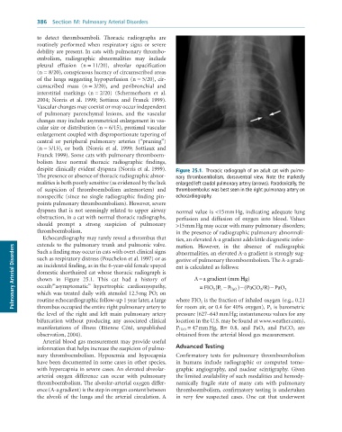
              despite clinically evident dyspnea (Norris et al. 1999).   Figure 25.1.  Thoracic	radiograph	of	an	adult	cat	with	pulmo-
              The presence or absence of thoracic radiographic abnor-  nary	 thromboembolism,	 dorsoventral	 view.	 Note	 the	 markedly
              malities is both poorly sensitive (as evidenced by the lack   enlarged	left	caudal	pulmonary	artery	(arrows).	Paradoxically,	the
              of  suspicion  of  thromboembolism  antemortem)  and   thromboembolus	was	best	seen	in	the	right	pulmonary	artery	on
              nonspecific (since no single radiographic finding pin-  echocardiography.
              points pulmonary thromboembolism). However, severe
              dyspnea that is not seemingly related to upper airway   normal  value  is  <15 mm Hg,  indicating  adequate  lung
              obstruction, in a cat with normal thoracic radiographs,   perfusion  and  diffusion  of  oxygen  into  blood.  Values
              should  prompt  a  strong  suspicion  of  pulmonary   >15 mm Hg may occur with many pulmonary disorders;
              thromboembolism.                                   in the presence of radiographic pulmonary abnormali-
                 Echocardiography may rarely reveal a thrombus that   ties, an elevated A-a gradient adds little diagnostic infor-
              extends  to  the  pulmonary  trunk  and  pulmonic  valve.
                                                                 mation.  However,  in  the  absence  of  radiographic
      Pulmonary Arterial Disorders  such as respiratory distress (Pouchelon et al. 1997) or as   gestive of pulmonary thromboembolism. The A-a gradi-
              Such a finding may occur in cats with overt clinical signs
                                                                 abnormalities, an elevated A-a gradient is strongly sug-
              an incidental finding, as in the 6-year-old female spayed
                                                                 ent is calculated as follows:
              domestic shorthaired cat whose thoracic radiograph is
                                                                         −
                                                                                    (
                                                                        A a gradient  mm Hg)
              shown  in  Figure  25.1.  This  cat  had  a  history  of
                                                                           FIO P b −
                                                                                    P H O −
                                                                                                   −
              occult/“asymptomatic”  hypertrophic  cardiomyopathy,
                                                                         =
                                                                               (
                                                                                       )
                                                                                          (PaCO /R) PaO 22
                                                                              2
                                                                                      2
              which was treated daily with atenolol 12.5 mg PO; on
              routine echocardiographic follow-up 1 year later, a large
                                                                 for room air, or 0.4 for 40% oxygen), P b is barometric
              thrombus occupied the entire right pulmonary artery to
                                                                 pressure (627–643 mm Hg; instantaneous values for any
              the  level  of  the  right  and  left  main  pulmonary  artery   where FIO 2  is the fraction of inhaled oxygen (e.g., 0.21
              bifurcation  without  producing  any  associated  clinical   location in the U.S. may be found at www.weather.com),
              manifestations  of  illness  (Etienne  Côté,  unpublished   P H2O  = 47 mm Hg,  R=  0.8,  and  PaO 2  and  PaCO 2   are
              observation, 2004).                                obtained from the arterial blood gas measurement.
                 Arterial blood gas measurement may provide useful
              information that helps increase the suspicion of pulmo-  Advanced Testing
              nary  thromboembolism.  Hypoxemia  and  hypocapnia   Confirmatory  tests  for  pulmonary  thromboembolism
              have been documented in some cases in other species,   in  humans  include  radiographic  or  computed  tomo-
              with hypercapnia in severe cases. An elevated alveolar-  graphic  angiography,  and  nuclear  scintigraphy.  Given
              arterial  oxygen  difference  can  occur  with  pulmonary   the limited availability of such modalities and hemody-
              thromboembolism. The alveolar-arterial oxygen differ-  namically  fragile  state  of  many  cats  with  pulmonary
              ence (A-a gradient) is the step in oxygen content between   thromboembolism, confirmatory testing is undertaken
              the alveoli of the lungs and the arterial circulation. A   in  very  few  suspected  cases.  One  cat  that  underwent

