
Page 545 - Feline diagnostic imaging
P. 545
558 31 Body Wall
(a)
(b) (c)
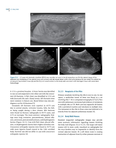
Figure 31.1 A 2-year-old domestic shorthair (DSH) was recently run over in the driveway by a car. On the lateral image of the
abdomen (a), thickening of the ventral body wall (arrows) and decreased detail within the subcutaneous fat are noted. On abdominal
ultrasound, hyperechoic tissue (b) and fluid (c) are noted ventral to the body wall consistent with damaged tissue with associated
hemorrhage.
8–12 in a proximal location. A hiatal hernia was identified 31.2.3 Neoplasia of the Ribs
in one cat and suspected in two other cats with the concur- Primary neoplasia involving the ribs is rare in cats. In one
rent rib fractures. A flail chest was identified in 1/12 cats
with fractured ribs and a hiatal hernia. Rib fractures were report, a multilobar tumor of bone was found in a cat
involving ribs 7–10 on computed tomography (CT) [7]. Two
more common in female cats. Renal failure may also pre-
dispose a cat for rib fractures [5]. cats with pulmonary carcinoma had evidence of metastasis
to multiple ribs on CT. Both cats had expansile rib lesions
Traumatic rib fractures were reported in 43/75 cats
due to animal attacks, vehicular trauma, falls, fan belt, with a periosteal reaction and osteolysis in multiple sites.
The metastasis to the ribs in these cases was believed to be
or being caught during a door closure. Rib fractures
were found on thoracic radiographs in 69/75 cases, and directly from the adjacent pulmonary masses [8].
6/75 on necropsy. The most common radiographic find-
ings were lung contusion, pneumothorax, pleural effu- 31.2.4 Body Wall Masses
sion, atelectasis, pneumomediastinum, and diaphragmatic Standard tangential radiographic images may provide
hernia (Figure 31.11). Cats with flail chest, pleural effu- some anatomic information regarding masses involving
sion, or diaphragmatic hernia had a higher mortality rate. the body wall (Figures 31.12–31.15). The larger soft tissue
Concurrent orthopedic injuries were found in 35/75 cats masses will be more easily visualized on radiographs but
with more injuries found cranial to the 13th vertebral the exact borders may be impossible to identify from the
body. Survival rate did not differ in cats with concurrent normal adjacent tissues. If a soft tissue mass is causing
orthopedic injuries [6]. destruction of adjacent bone by either pressure necrosis or