
Page 1138 - Veterinary Toxicology, Basic and Clinical Principles, 3rd Edition
P. 1138
1070 SECTION | XVI Feed and Water Contaminants
VetBooks.ir
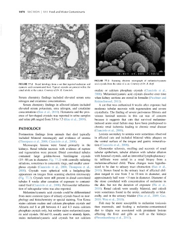
FIGURE 77.3 Scanning electron micrograph of melamine/cyanuric
FIGURE 77.2 Renal histology from a cat that ingested melamine and acid crystals from the urine of a cat. Courtesy of Dr. B. Hoff.
cyanuric acid contaminated food. Typical crystals are present within the
renal tubule at the center. Courtesy of Dr. R. Cianciolo. oxalate or calcium phosphate crystals (Cianciolo et al.,
2008). Melamine/cyanuric acid crystals dissolve over time
Serum chemistry findings included elevated serum urea when kidney sections are stored in formalin (Puschner and
nitrogen and creatinine concentrations. Reimschuessel, 2011).
Serum chemistry findings in affected infants included A cat that was euthanized 8 weeks after exposure had
elevated serum potassium, urea nitrogen, and creatinine moderate tubular necrosis with regeneration and severe
concentrations (Sun et al., 2010). Hematuria and the pres- crystalluria. The finding of severe perivenous fibrosis and
ence of fan-shaped crystals was reported in urine samples venous luminal stenosis in this cat was of concern
and urine pH ranged from 5.0 to 7.5 (Hau et al., 2009). because it suggests that cats that survived melamine-
induced acute renal failure may have been predisposed to
chronic renal ischemia leading to chronic renal disease
PATHOLOGY
(Cianciolo et al., 2008).
Postmortem findings from animals that died typically Lesions secondary to uremia were sometimes observed
included bilateral renomegaly and evidence of uremia in affected cats and included bilateral white plaques on
(Thompson et al., 2008; Cianciolo et al., 2008). the ventral surface of the tongue and gastric mineraliza-
Microscopic lesions were found primarily in the tion (Cianciolo et al., 2008).
kidneys. Renal tubular necrosis with evidence of rupture Glomerular sclerosis, swelling and necrosis of renal
and regeneration were present. Distal convoluted tubules tubular epithelium, tubular dilation with tubular dilation
contained large golden-brown birefringent crystals with lumenal crystals, and an interstitial lymphoplasmacy-
(15 80 μmin diameter; Fig. 77.2) with centrally radiating tic infiltrate were noted in a renal biopsy from a
striations, sometimes in concentric rings, and smaller amor- melamine-affected child. These changes were hypothe-
phous crystals (Cianciolo et al., 2008; Thompson et al., sized to be due to urinary tract obstruction (Sun et al.,
2008). Crystals were spherical with a hedgehog-like 2010). Stones found in the urinary tract of affected chil-
appearance on images from scanning electron microscopy dren ranged in size from 2 to 18 mm in diameter, and
(Fig. 77.3). Crystals were still present in distal convoluted approximately half were ,5 mm in diameter. Diameter of
tubules 8 weeks after cessation of exposure to contami- the stone correlated with concentration of melamine in
nated feed (Cianciolo et al., 2008). Perivascular inflamma- the diet, but not the duration of exposure (Hu et al.,
tion of subcapsular veins was also reported. 2010). Renal calculi were usually bilateral, and calculi
Melamine/cyanuric acid crystals could be differentiated were sometimes found in the ureter, unilaterally or bilat-
from more common types of urinary crystals based on mor- erally, and in the urinary bladder (Hau et al., 2009; Gao,
phology and histochemistry or special staining. Von Kossa 2010; Wen et al., 2010).
stains calcium oxalate and calcium phosphate crystals and Fish may be more susceptible to melamine toxicosis
Alizarin red S at pH between 4.1 and 4.3 stains calcium than mammals, and feeding a melamine-contaminated
phosphate crystals only, but neither stains melamine/cyanu- diet to tilapia was associated with prominent lesions
ric acid crystals. Oil red O, usually used to identify lipids, affecting the liver and gills as well as the kidneys
stains melamine/cyanuric acid crystals but not calcium (Phromkunthong et al., 2013).