
Page 916 - Veterinary Toxicology, Basic and Clinical Principles, 3rd Edition
P. 916
Poisonous Plants of the United States Chapter | 61 871
VetBooks.ir RO H C O O R HO H C O O R
C
C
2
2
R = Liver DNA
(toxicity)
P450
Liver
N activation N
R = Glutathione
PA Toxic
pyrrole
Urinary
excretion
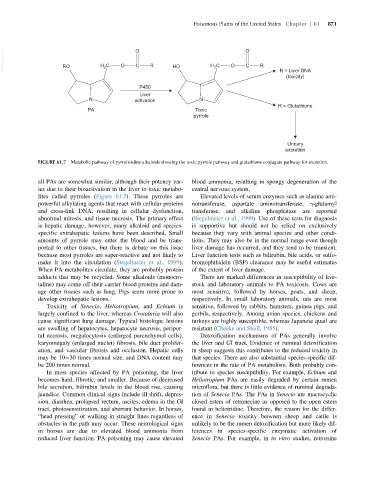
FIGURE 61.7 Metabolic pathway of pyrrolizidine alkaloids showing the toxic pyrrole pathway and glutathione conjugate pathway for excretion.
all PAs are somewhat similar, although their potency var- blood ammonia, resulting in spongy degeneration of the
ies due to their bioactivation in the liver to toxic metabo- central nervous system.
lites called pyrroles (Figure 61.7). These pyrroles are Elevated levels of serum enzymes such as alanine ami-
powerful alkylating agents that react with cellular proteins notransferase, aspartate aminotransferase, γ-glutamyl
and cross-link DNA, resulting in cellular dysfunction, transferase, and alkaline phosphatase are reported
abnormal mitosis, and tissue necrosis. The primary effect (Stegelmeier et al., 1999). Use of these tests for diagnosis
is hepatic damage; however, many alkaloid and species- is supportive but should not be relied on exclusively
specific extrahepatic lesions have been described. Small because they vary with animal species and other condi-
amounts of pyrrole may enter the blood and be trans- tions. They may also be in the normal range even though
ported to other tissues, but there is debate on this issue liver damage has occurred, and they tend to be transient.
because most pyrroles are super-reactive and not likely to Liver function tests such as bilirubin, bile acids, or sulfo-
make it into the circulation (Stegelmeier et al., 1999). bromophthalein (BSP) clearance may be useful estimates
When PA metabolites circulate, they are probably protein of the extent of liver damage.
adducts that may be recycled. Some alkaloids (monocro- There are marked differences in susceptibility of live-
taline) may come off their carrier blood proteins and dam- stock and laboratory animals to PA toxicosis. Cows are
age other tissues such as lung. Pigs seem more prone to most sensitive, followed by horses, goats, and sheep,
develop extrahepatic lesions. respectively. In small laboratory animals, rats are most
Toxicity of Senecio, Heliotropium, and Echium is sensitive, followed by rabbits, hamsters, guinea pigs, and
largely confined to the liver, whereas Crotalaria will also gerbils, respectively. Among avian species, chickens and
cause significant lung damage. Typical histologic lesions turkeys are highly susceptible, whereas Japanese quail are
are swelling of hepatocytes, hepatocyte necrosis, peripor- resistant (Cheeke and Shull, 1985).
tal necrosis, megalocytosis (enlarged parenchymal cells), Detoxification mechanisms of PAs generally involve
karyomegaly (enlarged nuclei) fibrosis, bile duct prolifer- the liver and GI tract. Evidence of ruminal detoxification
ation, and vascular fibrosis and occlusion. Hepatic cells in sheep suggests this contributes to the reduced toxicity in
may be 10 30 times normal size, and DNA content may that species. There are also substantial species-specific dif-
be 200 times normal. ferences in the rate of PA metabolism. Both probably con-
In most species affected by PA poisoning, the liver tribute to species susceptibility. For example, Echium and
becomes hard, fibrotic, and smaller. Because of decreased Heliotropium PAs are easily degraded by certain rumen
bile secretion, bilirubin levels in the blood rise, causing microflora, but there is little evidence of ruminal degrada-
jaundice. Common clinical signs include ill thrift, depres- tion of Senecio PAs. The PAs in Senecio are macrocyclic
sion, diarrhea, prolapsed rectum, ascites, edema in the GI closed esters of retronecine as opposed to the open esters
tract, photosensitization, and aberrant behavior. In horses, found in heliotridine. Therefore, the reason for the differ-
“head pressing” or walking in straight lines regardless of ence in Senecio toxicity between sheep and cattle is
obstacles in the path may occur. These neurological signs unlikely to be the rumen detoxification but more likely dif-
in horses are due to elevated blood ammonia from ferences in species-specific enzymatic activation of
reduced liver function. PA poisoning may cause elevated Senecio PAs. For example, in in vitro studies, retrorsine