
Page 1097 - Small Animal Internal Medicine, 6th Edition
P. 1097
CHAPTER 59 Diagnostic Tests for Nervous System and Neuromuscular Disorders 1069
VetBooks.ir
A
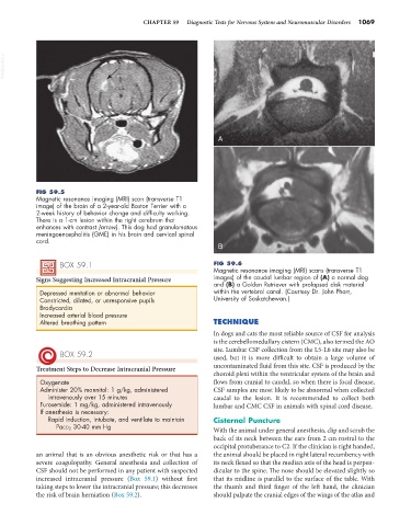
FIG 59.5
Magnetic resonance imaging (MRI) scan (transverse T1
image) of the brain of a 2-year-old Boston Terrier with a
2-week history of behavior change and difficulty walking.
There is a 1-cm lesion within the right cerebrum that
enhances with contrast (arrow). This dog had granulomatous
meningoencephalitis (GME) in his brain and cervical spinal
cord.
B
BOX 59.1 FIG 59.6
Magnetic resonance imaging (MRI) scans (transverse T1
Signs Suggesting Increased Intracranial Pressure images) of the caudal lumbar region of (A) a normal dog
and (B) a Golden Retriever with prolapsed disk material
Depressed mentation or abnormal behavior within the vertebral canal. (Courtesy Dr. John Pharr,
Constricted, dilated, or unresponsive pupils University of Saskatchewan.)
Bradycardia
Increased arterial blood pressure
Altered breathing pattern TECHNIQUE
In dogs and cats the most reliable source of CSF for analysis
is the cerebellomedullary cistern (CMC), also termed the AO
site. Lumbar CSF collection from the L5-L6 site may also be
BOX 59.2 used, but it is more difficult to obtain a large volume of
Treatment Steps to Decrease Intracranial Pressure uncontaminated fluid from this site. CSF is produced by the
choroid plexi within the ventricular system of the brain and
Oxygenate flows from cranial to caudal, so when there is focal disease,
Administer 20% mannitol: 1 g/kg, administered CSF samples are most likely to be abnormal when collected
intravenously over 15 minutes caudal to the lesion. It is recommended to collect both
Furosemide: 1 mg/kg, administered intravenously lumbar and CMC CSF in animals with spinal cord disease.
If anesthesia is necessary:
Rapid induction, intubate, and ventilate to maintain Cisternal Puncture
PaCO 2 30-40 mm Hg
With the animal under general anesthesia, clip and scrub the
back of its neck between the ears from 2 cm rostral to the
occipital protuberance to C2. If the clinician is right handed,
an animal that is an obvious anesthetic risk or that has a the animal should be placed in right lateral recumbency with
severe coagulopathy. General anesthesia and collection of its neck flexed so that the median axis of the head is perpen-
CSF should not be performed in any patient with suspected dicular to the spine. The nose should be elevated slightly so
increased intracranial pressure (Box 59.1) without first that its midline is parallel to the surface of the table. With
taking steps to lower the intracranial pressure; this decreases the thumb and third finger of the left hand, the clinician
the risk of brain herniation (Box 59.2). should palpate the cranial edges of the wings of the atlas and