
Page 1149 - Small Animal Internal Medicine, 6th Edition
P. 1149
CHAPTER 64 Encephalitis, Myelitis, and Meningitis 1121
made antemortem based on the typical MRI and CSF find- most dogs improve with treatment, the prognosis for perma-
ings and elimination of infectious and neoplastic diseases on nent recovery is poor. Dogs with forebrain signs have a sig-
VetBooks.ir the list of likely differentials. nificantly longer survival than dogs with multifocal or
Glucocorticoids can temporarily halt or reverse the pro-
brainstem signs, but dogs with seizures or MRI evidence of
gression of clinical signs in dogs with GME, particularly in
tocols recommend monitoring of MRI images and CSF
animals with slowly progressive signs associated with focal cerebral edema have limited survival. Some treatment pro-
disease. Clinical signs often recur quickly, with the median cytology before tapering drug doses, but that is rarely per-
survival time highly variable (7-114 days) depending on type formed. Radiation therapy has been reported to benefit some
and location of disease. More prolonged improvement in dogs with focal intracranial masses resulting from GME, and
clinical signs and survival can be seen when more aggres- results are similar to chemotherapy.
sive chemotherapy protocols are used, with median survival
times longer than 12 months expected when dogs with focal NECROTIZING MENINGOENCEPHALITIS
disease are treated with combinations of immunosuppressive NME is a breed-specific idiopathic inflammatory condition
drugs. Recommended drugs and protocols are outlined in affecting the brain of Pugs (Pug encephalitis) and Maltese
Box 64.3. Terriers. It has also been seen sporadically in the West High-
Comparative efficacy between protocols is difficult to land White Terrier, Chihuahua, Pekingese, Shih Tzu, Papil-
assess because of disease and patient variability and the lon, Coton du Tulear, and Lhasa Apso. Affected dogs first
failure to obtain a definitive pretreatment diagnosis. Dogs show clinical signs between 9 months and 7 years of age, with
with GME or MUE in the author’s hospital are usually treated a mean age of onset about 18 months in Pugs and 29 months
with a combination of prednisone, 4 cycles of cytosine ara- in other breeds. Female Pugs may be predisposed.
binoside, and either cyclosporine or azathioprine. Although Most dogs with NME are presented with an acute onset
of seizures and neurologic signs referable to the cerebrum
and meninges. They may have difficulty walking or may be
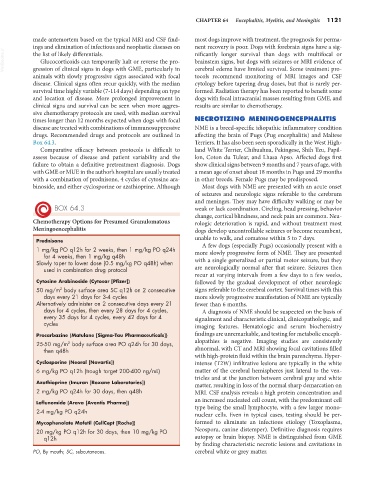
BOX 64.3 weak or lack coordination. Circling, head pressing, behavior
change, cortical blindness, and neck pain are common. Neu-
Chemotherapy Options for Presumed Granulomatous rologic deterioration is rapid, and without treatment most
Meningoencephalitis dogs develop uncontrollable seizures or become recumbent,
unable to walk, and comatose within 5 to 7 days.
Prednisone A few dogs (especially Pugs) occasionally present with a
1 mg/kg PO q12h for 2 weeks, then 1 mg/kg PO q24h more slowly progressive form of NME. They are presented
for 4 weeks, then 1 mg/kg q48h
Slowly taper to lower dose (0.5 mg/kg PO q48h) when with a single generalized or partial motor seizure, but they
used in combination drug protocol are neurologically normal after that seizure. Seizures then
recur at varying intervals from a few days to a few weeks,
Cytosine Arabinoside (Cytosar [Pfizer]) followed by the gradual development of other neurologic
2
50 mg/m body surface area SC q12h on 2 consecutive signs referable to the cerebral cortex. Survival times with this
days every 21 days for 3-4 cycles more slowly progressive manifestation of NME are typically
Alternatively administer on 2 consecutive days every 21 fewer than 6 months.
days for 4 cycles, then every 28 days for 4 cycles, A diagnosis of NME should be suspected on the basis of
every 35 days for 4 cycles, every 42 days for 4 signalment and characteristic clinical, clinicopathologic, and
cycles imaging features. Hematologic and serum biochemistry
Procarbazine (Matulane [Sigma-Tau Pharmaceuticals]) findings are unremarkable, and testing for metabolic enceph-
2
25-50 mg/m body surface area PO q24h for 30 days, alopathies is negative. Imaging studies are consistently
then q48h abnormal, with CT and MRI showing focal cavitations filled
with high-protein fluid within the brain parenchyma. Hyper-
Cyclosporine (Neoral [Novartis]) intense (T2W) infiltrative lesions are typically in the white
6 mg/kg PO q12h (trough target 200-400 ng/mL) matter of the cerebral hemispheres just lateral to the ven-
tricles and at the junction between cerebral gray and white
Azathioprine (Imuran [Roxane Laboratories]) matter, resulting in loss of the normal sharp demarcation on
2 mg/kg PO q24h for 30 days, then q48h MRI. CSF analysis reveals a high protein concentration and
an increased nucleated cell count, with the predominant cell
Leflunomide (Arava [Aventis Pharma]) type being the small lymphocyte, with a few larger mono-
2-4 mg/kg PO q24h nuclear cells. Even in typical cases, testing should be per-
Mycophenolate Mofetil (CellCept [Roche]) formed to eliminate an infectious etiology (Toxoplasma,
20 mg/kg PO q12h for 30 days, then 10 mg/kg PO Neospora, canine distemper). Definitive diagnosis requires
q12h autopsy or brain biopsy. NME is distinguished from GME
by finding characteristic necrotic lesions and cavitations in
PO, By mouth; SC, subcutaneous. cerebral white or grey matter.