
Page 1178 - Small Animal Internal Medicine, 6th Edition
P. 1178
1150 PART IX Nervous System and Neuromuscular Disorders
caused by tumor, diskospondylitis, synovial cyst, vertebral or
sacral osteochondrosis, or congenital bony malformations.
VetBooks.ir ity are all factors proposed to cause increased mechanical
Genetic predisposition, conformation, and physical activ-
stress on the IVD at the lumbosacral junction, promoting
type II disk prolapse at this site. Loss of the structural
strength of the disk worsens instability at the site, resulting
in proliferative changes in the articular facets, joint capsules,
and ligamentum flavum. Proliferative changes result in
further narrowing of the vertebral canal, compression of the
cauda equina, and compression of the nerve roots as they
exit the foramina (degenerative lumbosacral stenosis).
Clinical Features A
Compression of the nerve roots of the cauda equina results
in a very characteristic constellation of clinical signs. Affected
dogs are slow to rise from a prone position and reluctant to
run, sit up, jump, or climb stairs. Rear limb lameness worsens
with exercise as the blood vessels accompanying the spinal
nerve roots within the already crowded intervertebral
foramen dilate and further compress the nerve roots. Affected
dogs may be reluctant to raise or wag their tails.
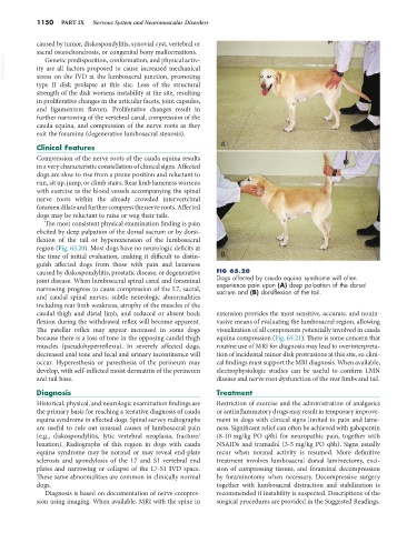
The most consistent physical examination finding is pain
elicited by deep palpation of the dorsal sacrum or by dorsi-
flexion of the tail or hyperextension of the lumbosacral
region (Fig. 65.20). Most dogs have no neurologic deficits at
the time of initial evaluation, making it difficult to distin- B
guish affected dogs from those with pain and lameness
caused by diskospondylitis, prostatic disease, or degenerative FIG 65.20
joint disease. When lumbosacral spinal canal and foraminal Dogs affected by cauda equina syndrome will often
experience pain upon (A) deep palpation of the dorsal
narrowing progress to cause compression of the L7, sacral, sacrum and (B) dorsiflexion of the tail.
and caudal spinal nerves, subtle neurologic abnormalities
including rear limb weakness, atrophy of the muscles of the
caudal thigh and distal limb, and reduced or absent hock extension provides the most sensitive, accurate, and nonin-
flexion during the withdrawal reflex will become apparent. vasive means of evaluating the lumbosacral region, allowing
The patellar reflex may appear increased in some dogs visualization of all components potentially involved in cauda
because there is a loss of tone in the opposing caudal thigh equina compression (Fig. 65.21). There is some concern that
muscles (pseudohyperreflexia). In severely affected dogs, routine use of MRI for diagnosis may lead to overinterpreta-
decreased anal tone and fecal and urinary incontinence will tion of incidental minor disk protrusions at this site, so clini-
occur. Hyperesthesia or paresthesia of the perineum may cal findings must support the MRI diagnosis. When available,
develop, with self-inflicted moist dermatitis of the perineum electrophysiologic studies can be useful to confirm LMN
and tail base. disease and nerve root dysfunction of the rear limbs and tail.
Diagnosis Treatment
Historical, physical, and neurologic examination findings are Restriction of exercise and the administration of analgesics
the primary basis for reaching a tentative diagnosis of cauda or antiinflammatory drugs may result in temporary improve-
equina syndrome in affected dogs. Spinal survey radiographs ment in dogs with clinical signs limited to pain and lame-
are useful to rule out unusual causes of lumbosacral pain ness. Significant relief can often be achieved with gabapentin
(e.g., diskospondylitis, lytic vertebral neoplasia, fracture/ (8-10 mg/kg PO q8h) for neuropathic pain, together with
luxation). Radiographs of this region in dogs with cauda NSAIDs and tramadol (3-5 mg/kg PO q8h). Signs usually
equina syndrome may be normal or may reveal end-plate recur when normal activity is resumed. More definitive
sclerosis and spondylosis of the L7 and S1 vertebral end treatment involves lumbosacral dorsal laminectomy, exci-
plates and narrowing or collapse of the L7-S1 IVD space. sion of compressing tissues, and foraminal decompression
These same abnormalities are common in clinically normal by foraminotomy when necessary. Decompressive surgery
dogs. together with lumbosacral distraction and stabilization is
Diagnosis is based on documentation of nerve compres- recommended if instability is suspected. Descriptions of the
sion using imaging. When available, MRI with the spine in surgical procedures are provided in the Suggested Readings.