Page 1261 - Small Animal Internal Medicine, 6th Edition
P. 1261
CHAPTER 73   Common Immune-Mediated Diseases   1233


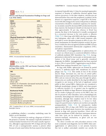
                   BOX 73.3                                      to respond (typically takes 3-5 days for maximal regenerative
                                                                 response)  or  because  of  antibodies  directed  against  bone
  VetBooks.ir  History and Physical Examination Findings in Dogs and   marrow precursors. In the latter situation, reticulocytes are
                                                                 destroyed before they enter the peripheral circulation. In the
            Cats With IMHA
             DOGS                           CATS                 absence of a regenerative response, a rapid fall in the hema-
                                                                 tocrit with little change in the serum total protein or albumin
             History                                             concentration should be considered suspicious for hemoly-
             Lethargy                       Lethargy             sis. In anemia caused by decreased RBC production from the
             Anorexia                       Anorexia
             Pallor                         Pallor               bone marrow, the hematocrit should not decrease by more
             Icterus                        Icterus              than approximately 1% per day, whereas in blood loss
             Vomiting                       Vomiting             anemia, the drop in the hematocrit is usually accompanied
             Collapse                       Pica                 by a concurrent decrease in the  total protein or  albumin
             Weakness                                            (Table 73.1). Most dogs with IMHA also have an inflamma-
             Physical Examination (Additional Findings)
             Systolic heart murmur          Systolic heart murmur  tory leukogram, often with a shift toward immature cells,
             Pyrexia                        Pyrexia              and mild to severe thrombocytopenia in 60% of cases. Pro-
             Tachycardia                    Hypothermia          posed mechanisms for thrombocytopenia include the pres-
             Tachypnea                      Lymphadenomegaly     ence of antibodies directed against platelets and RBCs (Evans
             Pallor                         Pallor               syndrome), disseminated intravascular coagulation (DIC),
             Icterus                        Icterus              and splenic sequestration.
             Splenomegaly
             Hepatomegaly                                          Identification of autoagglutination or spherocytosis (2+
             Abdominal pain                                      or more) on a blood smear is considered definitive evidence
                                                                 of  antibody-mediated  RBC hemolysis (Fig. 73.3).  Autoag-
            IMHA, Immune-mediated hemolytic anemia.              glutination is detected by macroscopic or microscopic exam-
                                                                 ination of the blood smear and is generally considered
                                                                 diagnostic for IMHA. Autoagglutination has been reported
                   BOX 73.4                                      in up to 78% of canine IMHA. Agglutination must be distin-

            Abnormalities on the CBC and Serum Chemistry Profile   guished from rouleaux formation (see Chapter 82).
            in Dogs With IMHA                                      In dogs with IMHA, spherocytes are formed by partial
                                                                 removal of antibody-coated RBC membranes by macro-
             CBC                                                 phages (see  Fig. 73.2). This results in loss of the normal
             Anemia                                              discoid shape, decreased size, and loss of central pallor.
             Polychromasia                                       Spherocytes are more rigid and less deformable than normal
             Autoagglutination                                   RBCs and are removed when they pass through the spleen.
             Spherocytosis                                       Spherocytes are readily identified in the dog but difficult to
             Ghost cells                                         recognize in cats because of the lack of significant central
             Evidence of inflammation (increased neutrophils, bands,   pallor in their normal RBCs. Spherocytes are considered a
               metamyelocytes, monocytes)                        hallmark morphologic change in IMHA, and when present
             Thrombocytopenia                                    in sufficient numbers (2+ or greater) may be regarded as

             Biochemical Profile                                 diagnostic for IMHA in dogs. However, because spherocytes
             Hemoglobinemia                                      are the results of phagocytic activity on the RBCs, they can
             Hemoglobinuria                                      also be present in other disorders, such as hemophagocytic
             Hyperbilirubinemia                                  syndrome, hemophagocytic histiocytosis, and zinc-induced
             Hyperbilirubinuria                                  hemolysis, although their numbers tend to be low when
             Increased alanine aminotransferase                  compared with dogs with IMHA (i.e., 1+ versus 2+). Tech-
             Increased alkaline phosphatase                      niques for quantification of spherocyte numbers are typically
                                                                 semiquantitative (Table 73.2). In retrospective studies
            CBC, Complete blood cell count; IMHA, immune-mediated   approximately 90% of dogs with IMHA have spherocytes
            hemolytic anemia.
                                                                 present on the blood smear; however, low numbers may be
                                                                 present in dogs with peracute hemolysis. Ghost cells are
            at establishing whether a secondary underlying cause for   remnant membranes of RBCs that have undergone intravas-
            IMHA can be identified.                              cular lysis. Lysis can be induced by immune- or non–
              The first requirement for making a diagnosis of IMHA is   immune-mediated mechanisms, so ghost cells are not
            the presence of anemia. The anemia is typically moderate to   diagnostic for IMHA.
            marked (median hematocrit of 13%) and is usually regenera-  The direct Coombs test with polyvalent antisera is the
            tive, although in approximately 30% of dogs and more than   most commonly used diagnostic test for IMHA when auto-
            50% of cats a nonregenerative anemia is present either   agglutination or spherocytosis is not present; however,
            because of acute onset before the bone marrow has had time   this test is neither particularly sensitive nor specific for

