
Page 206 - Small Animal Internal Medicine, 6th Edition
P. 206
178 PART I Cardiovascular System Disorders
inspiration and expiration is usually greater than 10 mm Hg in patients with moderate to large pericardial effusions. Lung
in patients with cardiac tamponade and pulsus paradoxus. sounds are muffled over the ventral thorax in those with
VetBooks.ir Pulsus paradoxus is not always discernible by femoral pulse pleural effusion. Although pericardial effusion does not
cause a murmur, concurrent cardiac disease may do so.
palpation.
Clinical Features Infectious pericarditis may be accompanied by fever; rarely,
a pericardial friction rub may be heard.
Clinical findings in patients with cardiac tamponade usually
reflect right-sided CHF and poor cardiac output. Before Diagnosis
obvious ascites develop, nonspecific signs might include Clinicopathologic findings
lethargy, weakness, poor exercise tolerance, and inappetence Hematologic and biochemical test results generally are
or other GI signs. In many cases, the client history describes nonspecific. The complete blood count (CBC) might indicate
complaints of exercise intolerance, abdominal enlargement, mild nonregenerative anemia, especially in patients with
tachypnea or difficulty breathing, collapse, and sometimes neoplastic disease, or may suggest inflammation or infection.
cough or vomiting. However, rapid pericardial fluid accu- Cardiac HSA can be associated with a regenerative anemia,
mulation can cause acute tamponade, shock, and death increased numbers of nucleated red blood cells and schisto-
without signs of ascites, pleural effusion, or marked radio- cytes (with or without acanthocytes), and thrombocytope-
graphic cardiomegaly. Pulmonary edema, jugular venous nia. Mild hypoproteinemia is seen in some patients. Mild
distension, and hypotension might be evident in such cases, increases in liver enzyme activities and prerenal azotemia
though. A history of collapse could be more common in can occur secondary to hepatic congestion and poor cardiac
dogs with neoplastic disease. Some cases with long-standing output. More pronounced liver enzyme elevation sometimes
disease develop marked loss of lean body mass (cachexia; occurs with neoplastic effusions. Other biochemical abnor-
Fig. 9.3). malities that have been reported in dogs with pericardial
Jugular vein distention or a positive hepatojugular reflux, effusion include hyperlactatemia, hyponatremia, hyperglyce-
hepatomegaly, ascites, labored respirations, and weak femoral mia, and hypermagnesemia. Pleural and peritoneal fluids in
pulses are common physical examination findings. Pleural dogs and cats with cardiac tamponade are usually modified
effusion and ascites also occur in both cats and dogs with transudates.
cardiac tamponade. Ascites may be more prevalent in dogs Circulating cardiac troponin (cTnI) concentration can
without an identifiable mass lesion, reflecting a more gradual increase as a result of ischemia or myocardial invasion. An
worsening or progression of tamponade. A palpable decrease elevated cTnI helps differentiate pericardial effusion caused
in arterial pulse strength during inspiration (pulsus para- by HSA from other causes, especially in cases where a mass
doxus) might be discernible in some dogs with tamponade. lesion is not obvious on echocardiogram. HSA that does
Sinus tachycardia, pale mucous membranes, and prolonged not affect the heart does not increase cTnI concentration.
capillary refill time are common, as manifestations of high Although pericardial fluid also can be used for cTnI mea-
sympathetic tone. The precordial impulse is weak when the surement, this does not provide improved sensitivity. Serum
pericardial fluid volume is large. Heart sounds are muffled NT-proBNP concentration is likely to be low in patients
with pericardial effusion, in contrast to other cardiac
diseases.
RADIOGRAPHY
Pericardial effusion enlarges the cardiac silhouette (Fig. 9.4).
A massive amount of pericardial fluid causes the classic
globoid-shaped heart shadow on both radiographic views.
Nevertheless, radiographic vertebral heart score (VHS) or
measures of sphericity are only moderately accurate for
differentiating pericardial effusion from other cardiac dis-
orders. These indices are not sensitive or specific enough
to reliably identify dogs with pericardial effusion and
cardiac tamponade from other causes of right-sided CHF
signs. Smaller fluid volumes allow various cardiac contours
to be identified, especially dorsally. Other findings asso-
ciated with tamponade (as well as other causes of right-
sided CHF) include pleural effusion, a distended caudal
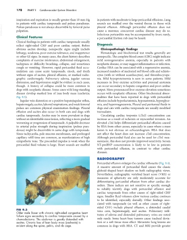
FIG 9.3 vena cava, hepatomegaly, and ascites. Pulmonary infil-
Older male Boxer with chronic right-sided congestive heart
failure signs secondary to cardiac tamponade caused by a trates of edema and distended pulmonary veins are noted
chemodectoma. The abdomen is greatly distended with only rarely. Some heart-base tumors cause tracheal devia-
ascites. Chronic loss of lean body mass (cachexia) is tion or a soft tissue mass effect. Metastatic lung lesions are
evident along the spine, pelvis, and rib cage. common in dogs with HSA. CT and MRI provide greater