
Page 224 - Small Animal Internal Medicine, 6th Edition
P. 224
196 PART I Cardiovascular System Disorders
old, HWD is also diagnosed in dogs younger than 1 year (but As with other causes of PAH, a loud and often split-second
older than 6 months) of age, as well as in geriatric animals. heart sound (S 2 ) and a murmur of TR are commonly heard
VetBooks.ir Males are affected two to four times as often as females. on auscultation. Severe pulmonary arteritis and thrombo-
embolism (see p. 224) can lead to marked dyspnea with
Large-breed dogs and those living mainly outdoors are at
cyanosis, hemoptysis, fever, DIC, thrombocytopenia, and
much greater risk of infection than small-breed and indoor
dogs. The length of the haircoat does not appear to affect epistaxis. These signs, as well as anemia and hemoglobinuria,
infection risk. also are associated with caval syndrome (see p. 202). Aber-
Dogs diagnosed by a positive routine screening test often rant worm migration to the central nervous system, eye,
are asymptomatic. Dogs with occult disease and those not femoral arteries, subcutis, peritoneal cavity, and other sites
routinely tested are more likely to have advanced pulmonary causes signs related to the organ involved.
arterial changes and clinical signs associated with pulmo-
nary hypertension, eosinophilic pneumonitis, and secondary Diagnosis
right-sided cardiac remodeling. Dogs with clinical disease
generally have a history of poor exercise tolerance, cough, RADIOGRAPHY
respiratory difficulty, syncope, weight loss, or abdominal dis- Radiographic findings can be normal early in the disease
tension with fluid. Overall severity of HWD is classified process or with only a few worms. However, marked changes
based on severity of clinical signs, radiographic signs, and develop rapidly in dogs with heavy worm burdens. Charac-
clinicopathologic abnormalities (Table 10.2). Of all dogs teristic findings are those suggestive of severe PAH, includ-
diagnosed with HWD, the majority (70%) are asymptomatic ing RV enlargement, a pulmonary trunk bulge, and centrally
(Class 1), approximately 25% have exercise intolerance or enlarged and tortuous lobar pulmonary arteries with periph-
respiratory signs secondary to pneumonitis or pulmonary eral blunting (Fig. 10.1 and p. 191). Patchy pulmonary inter-
hypertension (Class 2), and less than 5% present with right- stitial or alveolar infiltrates suggestive of pneumonitis, PTE,
sided CHF or caval syndrome (Class 3 or 4). or fibrosis are also common, especially in the caudal lobes.
Physical examination findings are often normal in dogs These pulmonary opacities may be mainly perivascular.
with early or mild disease. However, severe disease is associ- Occasionally eosinophilic granulomatosis occurs, with more
ated with poor body condition, tachypnea or dyspnea, clini- organized interstitial nodules, enlarged bronchial lymph
cal signs of severe PAH (see p. 191), or evidence of right-sided nodes, and sometimes pleural effusion. In severe cases where
CHF (see p. 201). Increased or abnormal lung sounds PAH has progressed to cause right-sided CHF, caudal vena
(wheezes and crackles) can accompany pulmonary paren- cava enlargement, hepatosplenomegaly, and abdominal or
chymal involvement as seen with eosinophilic pneumonitis. pleural effusion may be seen.
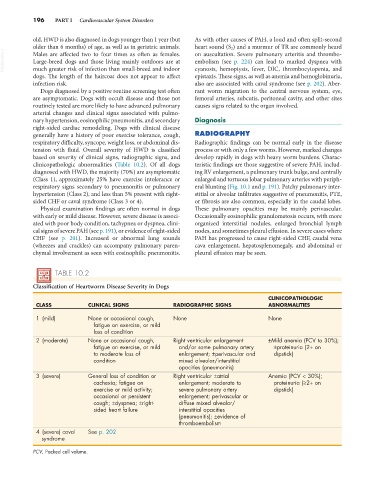
TABLE 10.2
Classification of Heartworm Disease Severity in Dogs
CLINICOPATHOLOGIC
CLASS CLINICAL SIGNS RADIOGRAPHIC SIGNS ABNORMALITIES
1 (mild) None or occasional cough, None None
fatigue on exercise, or mild
loss of condition
2 (moderate) None or occasional cough, Right ventricular enlargement ±Mild anemia (PCV to 30%);
fatigue on exercise, or mild and/or some pulmonary artery ±proteinuria (2+ on
to moderate loss of enlargement; ±perivascular and dipstick)
condition mixed alveolar/interstitial
opacities (pneumonitis)
3 (severe) General loss of condition or Right ventricular ±atrial Anemia (PCV < 30%);
cachexia; fatigue on enlargement; moderate to proteinuria (≥2+ on
exercise or mild activity; severe pulmonary artery dipstick)
occasional or persistent enlargement; perivascular or
cough; ±dyspnea; ±right- diffuse mixed alveolar/
sided heart failure interstitial opacities
(pneumonitis); ±evidence of
thromboembolism
4 (severe) caval See p. 202
syndrome
PCV, Packed cell volume.