
Page 948 - Small Animal Internal Medicine, 6th Edition
P. 948
920 PART VII Metabolic and Electrolyte Disorders
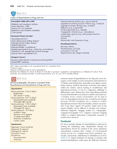
BOX 53.3
VetBooks.ir Causes of Hyperkalemia in Dogs and Cats Potassium-sparing diuretics (e.g., spironolactone)
Transcellular Shifts (ICF to ECF)
Metabolic and respiratory acidosis Angiotensin-converting enzyme inhibitors (e.g., enalapril)
Insulin deficiency—DKA Angiotensin-receptor blockers (e.g., losartan)
Acute tumor lysis syndrome β-Blockers (e.g., propranolol)
Reperfusion post–thrombus dissolution Cardiac glycosides (e.g., digitalis)
Crush injuries Prostaglandin inhibitors (e.g., indomethacin)
α-Adrenergic agonists (e.g., phenylpropanolamine)
Decreased Urinary Excretion Cyclosporine
Hypoadrenocorticism* Heparin
Acute oliguric-anuric kidney disease* Nonsteroidal antiinflammatory drugs
End-stage chronic kidney disease Pseudohyperkalemia
Urethral obstruction*
Ruptured bladder—uroabdomen* Hemolysis (Akita)
6
Selected gastroenteritis (e.g., trichuriasis, salmonellosis) Thrombocytosis (>10 /µL)
5
Chylothorax with repeated pleural fluid drainage Leukocytosis (>10 /µL)
Hyporeninemic hypoaldosteronism Hypernatremia (dry reagent methods)
Iatrogenic Causes†
Excessive administration of potassium-containing fluids*
Expired RBC transfusion
DKA, Diabetic ketoacidosis; ECF, extracellular fluid; ICF, intracellular fluid.
*Common causes.
†Require contributing factors to cause hyperkalemia.
Modified from DiBartola SP, Autran de Morais H: Disorders of potassium: hypokalemia and hyperkalemia. In DiBartola SP, editor: Fluid,
electrolyte, and acid-base disorders in small animal practice, ed 4, St Louis, 2012, Saunders Elsevier.
BOX 53.4 common causes of hyperkalemia in the dog and cat are iat-
rogenic, most notably excessive potassium administration in
Electrocardiographic Alterations Associated With IV fluids; renal dysfunction, especially acute oliguric-anuric
Hyperkalemia and Hypokalemia in Dogs and Cats kidney disease, urethral obstruction (tomcats), and rupture
within the urinary system leading to uroabdomen; and
Hyperkalemia hypoadrenocorticism. It can be a diagnostic challenge to
Serum potassium: 5.6-6.5 mEq/L differentiate renal dysfunction from hypoadrenocorticism
Bradycardia because both disorders can result in a similar clinical picture.
Tall, narrow T waves
Serum potassium: 6.6-7.5 mEq/L A baseline serum cortisol concentration can be used to
Decreased R-wave amplitude rule out hypoadrenocorticism, but an adrenocorticotropic
Prolonged QRS interval hormone (ACTH) stimulation test is needed to confirm
Serum potassium: 7.0-8.5 mEq/L hypoadrenocorticism when the baseline cortisol concentra-
Decreased P-wave amplitude tion is less than 2 µg/dL (55 nmol/L). Small rents in the
Prolonged P-R interval urinary bladder can be difficult to identify, and contrast-
Serum potassium: >8.5 mEq/L enhanced diagnostic imaging studies (i.e., radiographic,
Invisible P wave computed tomography [CT], magnetic resonance imaging
Deviation of ST segment [MRI]) or surgical exploration is frequently necessary to
Complete heart block confirm their presence.
Ventricular arrhythmias
Cardiac arrest Treatment
Hypokalemia For most animals, therapy for hyperkalemia is directed at
Depressed T-wave amplitude treating the underlying cause. Symptomatic therapy for
Depressed ST segment hyperkalemia should be initiated if the serum potassium
Prolonged QT interval concentration is greater than 7 mEq/L, or if pronounced
Prominent U wave cardiac toxicity (i.e., complete heart block, premature ven-
Arrhythmias tricular contractions, arrhythmias) is identified on an ECG
Supraventricular (Table 53.2). Rapid institution of therapy in animals with
Ventricular marked hyperkalemia could mean the difference between