
Page 21 - Veterinary diagnostic imaging birds exotic pets wildlife
P. 21
CHAPTER 1 III Avian Radiography and Radiographic Diagnosis 17
A
B
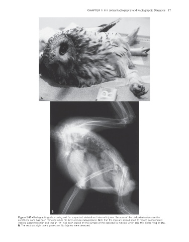
Figure 1-15 • Radiographing a burrowing owl for suspected skeletal and internal injuries. Because of the bird’s diminutive size the
anesthetic cone has been removed while the bird is being radiographed. Note that the legs are spread apart to reduce concentrated
visceral superimposition and that an “R” has been placed on the surface of the cassette to indicate which side the bird is lying on (A).
B, The resultant right lateral projection. No injuries were detected.
2/11/2008 10:50:48 AM
ch001-A02527.indd 17 2/11/2008 10:50:48 AM
ch001-A02527.indd 17