
Page 244 - Veterinary diagnostic imaging birds exotic pets wildlife
P. 244
240 SECTION II III The Mammals
III NORMAL RADIOGRAPHIC ANATOMY is disproportionally small when compared with the
much wider and longer abdomen. As seen in both
Little sets the guinea pig apart from the rest of the standard radiographic projections (Figure 21-11), the
small rodents, although this class of mammals does visible portion of the thorax comprises only about one-
have distinguishing morphological features. sixth of the total length of the torso.
Second, the thoracic viscera typically appear vague,
the result of the normally poor or at best mediocre
Skull
contrast between the heart and lung. Thoracic super-
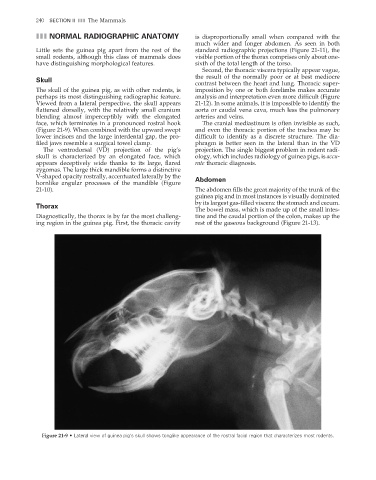
The skull of the guinea pig, as with other rodents, is imposition by one or both forelimbs makes accurate
perhaps its most distinguishing radiographic feature. analysis and interpretation even more diffi cult (Figure
Viewed from a lateral perspective, the skull appears 21-12). In some animals, it is impossible to identify the
flattened dorsally, with the relatively small cranium aorta or caudal vena cava, much less the pulmonary
blending almost imperceptibly with the elongated arteries and veins.
face, which terminates in a pronounced rostral hook The cranial mediastinum is often invisible as such,
(Figure 21-9). When combined with the upward swept and even the thoracic portion of the trachea may be
lower incisors and the large interdental gap, the pro- difficult to identify as a discrete structure. The dia-
fi led jaws resemble a surgical towel clamp. phragm is better seen in the lateral than in the VD
The ventrodorsal (VD) projection of the pig’s projection. The single biggest problem in rodent radi-
skull is characterized by an elongated face, which ology, which includes radiology of guinea pigs, is accu-
appears deceptively wide thanks to its large, fl ared rate thoracic diagnosis.
zygomas. The large thick mandible forms a distinctive
V-shaped opacity rostrally, accentuated laterally by the Abdomen
hornlike angular processes of the mandible (Figure
21-10). The abdomen fills the great majority of the trunk of the
guinea pig and in most instances is visually dominated
by its largest gas-filled viscera: the stomach and cecum.
Thorax
The bowel mass, which is made up of the small intes-
Diagnostically, the thorax is by far the most challeng- tine and the caudal portion of the colon, makes up the
ing region in the guinea pig. First, the thoracic cavity rest of the gaseous background (Figure 21-13).
Figure 21-9 • Lateral view of guinea pig’s skull shows tonglike appearance of the rostral facial region that characterizes most rodents.
2/11/2008 11:09:42 AM
ch021-A02527.indd 240
ch021-A02527.indd 240 2/11/2008 11:09:42 AM