
Page 27 - Veterinary diagnostic imaging birds exotic pets wildlife
P. 27
CHAPTER 1 III Avian Radiography and Radiographic Diagnosis 23
A
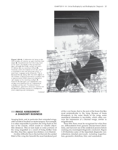
Figure 1-18 • A, A Halloween bat hangs in the
front window of a home, as seen from the front
steps. B, Viewed directly from the living room
of the home, the details of the bat become
clear, although the blinds, control rod, and a
Christmas star are overlying it. C and D,
However, when the shadow of the bat, which
is projected on the wall above the couch, is
examined, it appears quite differently. This is
because the bat, along with the other objects in
the window, is being projected on a surface that
is approximately 6 feet away and oriented at
right angles to the incoming sunlight. E,
Examine the bat again, as it appeared in the
window and subsequently as it appeared as
a shadow on the wall. The phenomena of
magnification, geometric distortion, and blur are
all relevant potential sources of misdiagnosis
when analyzing avian radiographs. B
Continued
III IMAGE ASSESSMENT: of the x-ray beam, that is, the part of the beam that lies
A SHADOWY BUSINESS most perpendicular to the wing. Because of beam
divergence in the outer thirds of the wing, some
anatomical distortion is inevitable, which often con-
Imaging birds, and in particular their extended wings, veys the impression of a blurred image, similar to
often results in less than accurate imagery. For example, magnifi cation.
it is often impossible to position the wing flush on the These fi lm fl aws must be recognized for what they
face of the cassette, which results in variable object- are, differentiated from genuine disease-based altera-
film distances. This in turn leads to some portions of tions, and factored out of the analytical process before
the wing magnified as a result of being farther from reaching any meaningful diagnostic conclusion. Figure
the fi lm, thus they appear less distinct, even blurred. 1-18 illustrates some of the important diagnostic con-
Another difficulty lies in the fact that only the central siderations detailed previously, including magnifi ca-
third of the wing lies beneath the most functional part tion, geometric distortion, blur, and summation.
2/11/2008 10:50:54 AM
ch001-A02527.indd 23
ch001-A02527.indd 23 2/11/2008 10:50:54 AM