
Page 330 - Veterinary diagnostic imaging birds exotic pets wildlife
P. 330
326 SECTION II III The Mammals
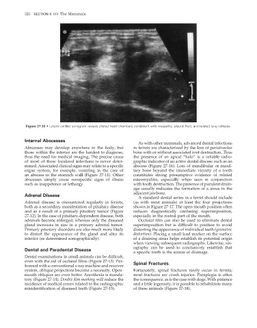
Figure 27-10 • Lateral cardiac sonogram reveals dilated heart chambers consistent with myopathy, pleural fluid, and related lung collapse.
Internal Abscesses
As with other mammals, advanced dental infections
Abscesses may develop anywhere in the body, but in ferrets are characterized by the loss of perialveolar
those within the interior are the hardest to diagnose, bone with or without associated root destruction. Thus
thus the need for medical imaging. The precise cause the presence of an apical “halo” is a reliable radio-
of most of these localized infections is never deter- graphic indicator of an active dental disease such as an
mined. Associated clinical signs may relate to a specifi c abscess (Figure 27-16). Loss of mandibular or maxil-
organ system, for example, vomiting in the case of lary bone beyond the immediate vicinity of a tooth
an abscess in the stomach wall (Figure 27-11). Other constitutes strong presumptive evidence of related
abscesses simply cause nonspecific signs of illness osteomyelitis, especially when seen in conjunction
such as inappetence or lethargy. with tooth destruction. The presence of purulent drain-
age usually indicates the formation of a sinus in the
adjacent jawbone.
Adrenal Disease
A standard dental series in a ferret should include
Adrenal disease is encountered regularly in ferrets, (as with most animals) at least the four projections
both as a secondary manifestation of pituitary disease shown in Figure 27-17. The open-mouth position often
and as a result of a primary pituitary tumor (Figure reduces diagnostically confusing superimposition,
27-12). In the case of pituitary-dependent disease, both especially in the rostral part of the mouth.
adrenals become enlarged, whereas only the diseased Occlusal film can also be used to eliminate dental
gland increases in size in a primary adrenal tumor. superimposition but is difficult to position to avoid
Primary pituitary disorders are also much more likely distorting the appearance of individual teeth (geometric
to distort the appearance of the gland and alter its distortion). Placing a small lead marker on the surface
interior (as determined sonographically). of a draining sinus helps establish its potential origin
when viewing subsequent radiographs. Likewise, sin-
ography can be used to conclusively establish that
Dental and Paradental Disease
a specifi c tooth is the source of drainage.
Dental examinations in small animals can be diffi cult,
even with the aid of occlusal films (Figure 27-13). Per- Spinal Fractures
formed with a conventional x-ray machine and receiver
system, oblique projections become a necessity. Open- Fortunately, spinal fractures rarely occur in ferrets;
mouth obliques are even better. Anesthesia is manda- most fractures are crush injuries. Paraplegia is often
tory (Figure 27-14). Double fi lm marking will reduce the the consequence, as is the case with dogs. With patience
incidence of medical errors related to the radiographic and a little ingenuity, it is possible to rehabilitate many
misidentifi cation of diseased teeth (Figure 27-15). of these animals (Figure 27-18).
2/11/2008 11:44:04 AM
ch027-A02527.indd 326 2/11/2008 11:44:04 AM
ch027-A02527.indd 326