
Page 592 - Withrow and MacEwen's Small Animal Clinical Oncology, 6th Edition
P. 592
570 PART IV Specific Malignancies in the Small Animal Patient
VetBooks.ir
A B
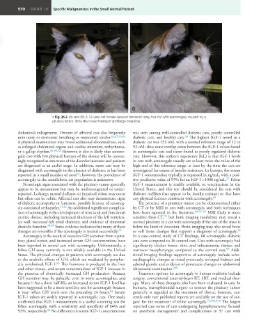
• Fig. 26.2 (A) and (B) A 12-year-old female spayed domestic long-hair cat with acromegaly caused by a
pituitary tumor. Note the broad forehead and large mandible.
abdominal enlargement. Owners of affected cats also frequently was seen among well-controlled diabetic cats, poorly controlled
note noisy or stertorous breathing or respiratory stridor. 86,87,90,91 diabetic cats, and healthy cats. The highest IGF-1 noted in a
90
A physical examination may reveal additional abnormalities, such diabetic cat was 153 nM, with a normal reference range of 12 to
as enlarged abdominal organs and cardiac murmurs, arrhythmias, 92 nM; thus some overlap exists between the IGF-1 values found
or a gallop rhythm. 81,86,92 However, it also is likely that acrome- in acromegalic cats and those found in poorly regulated diabetic
galic cats with few physical features of the disease will be increas- cats. However, this author’s experience (KL) is that IGF-1 levels
ingly recognized as awareness of the disorder increases and patients in cats with acromegaly usually are at least twice the value of the
are diagnosed at an earlier stage. In addition, more cats may be high end of this reference range, at least by the time the cats are
diagnosed with acromegaly in the absence of diabetes, as has been investigated for causes of insulin resistance. In Europe, the serum
reported in a small number of cases ; however, the prevalence of IGF-1 concentration typically is expressed in ng/mL, with a posi-
93
87
acromegaly in the nondiabetic cat population is unknown. tive predictive value of 95% for an IGF-1 >1000 ng/mL. Feline
Neurologic signs associated with the pituitary tumor generally IGF-1 measurement is readily available to veterinarians in the
appear to be uncommon but may be underrecognized or under- United States, and this test should be considered for cats with
reported. Lethargy, mental dullness, or impaired vision may occur diabetes mellitus that appear to be insulin resistant or that have
but often can be subtle. Affected cats also may demonstrate signs any physical features consistent with acromegaly. 89
of diabetic neuropathy or lameness, possibly because of acromeg- The presence of a pituitary tumor can be demonstrated either
aly-associated arthopathy. 81,86 An additional significant complica- by CT or by MRI in cats with acromegaly, and both techniques
tion of acromegaly is the development of structural and functional have been reported in the literature. 86,96–99 MRI likely is more
99
cardiac disease, including increased thickness of the left ventricu- sensitive than CT, but both imaging modalities may reveal a
lar wall, increased left atrial diameter, and evidence of abnormal normal pituitary in a cat with acromegaly if the size of the mass is
diastolic function. 94,95 Some evidence indicates that many of these below the limit of detection. Brain imaging may also reveal bone
98
changes are reversible if the acromegaly is treated successfully. 95 or soft tissue changes that support a diagnosis of acromegaly.
Acromegaly is the result of excessive GH secretion from a pitu- In a case-control study of CT findings, 68 acromegalic diabetic
itary gland tumor, and increased serum GH concentrations have cats were compared to 36 control cats. Cats with acromegaly had
been reported in several cats with acromegaly. Unfortunately, a significantly thicker bones, skin, and subcutaneous tissues, and
feline GH assay currently is not reliably available in the United narrower nasopharynges compared to the control cats. Addi-
99
States. The physical changes in patients with acromegaly are due tional imaging findings supportive of acromegaly include echo-
to the anabolic effects of GH, which are mediated by peripher- cardiographic changes as noted previously, enlarged kidneys and
83
ally synthesized IGF-1. This hormone is produced in the liver adrenal glands, and evidence of pancreatic changes on abdominal
and other tissues, and serum concentrations of IGF-1 increase in ultrasound examination. 100
the presence of chronically increased GH production. Because Treatment options for acromegaly in human medicine include
GH secretion may be pulsatile, even in some acromegalics, and surgery, conventional external-beam RT, SRT, and medical ther-
because it has a short half-life, an increased serum IGF-1 level has apy. Many of these therapies also have been evaluated in cats. In
been suggested to be a more sensitive test for acromegaly because humans, transsphenoidal surgery to remove the pituitary tumor
it may reflect GH levels over the preceding 24 hours. Serum generally is regarded as the treatment of choice; however, cur-
83
IGF-1 values are widely reported in acromegalic cats. One study rently only rare published reports are available on the use of sur-
confirmed that IGF-1 measurement is a useful screening test for gery for the treatment of feline acromegaly. 70,101,102 The largest
feline acromegaly, with a sensitivity and specificity of 84% and reported case series of cats undergoing hypophysectomy focused
92%, respectively. No difference in serum IGF-1 concentrations on anesthetic management and complications in 37 cats with
90