
Page 33 - Small Animal Internal Medicine, 6th Edition
P. 33
CHAPTER 1 Clinical Manifestations of Cardiac Disease 5
BOX 1.4
VetBooks.ir Abnormal Mucous Membrane Color
Pale Mucous Membranes
Anemia
Poor cardiac output/high sympathetic tone
Injected, Brick-Red Membranes
Polycythemia (erythrocytosis)
Sepsis
Excitement
Other causes of peripheral vasodilation
Cyanotic Mucous Membranes*
Pulmonary parenchymal disease
Airway obstruction
Pleural space disease
Pulmonary edema
Right-to-left shunting congenital cardiac defect
Hypoventilation
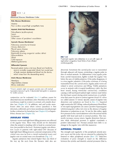
Shock FIG 1.4
Cold exposure Prominent jugular vein distention in a cat with signs of
Methemoglobinemia right-sided congestive heart failure from dilated
cardiomyopathy.
Differential Cyanosis
Reversed patent ductus arteriosus (head and forelimbs
receive normally oxygenated blood, but caudal part abnormal. Sometimes the carotid pulse wave is transmitted
of body receives desaturated blood via the ductus, through adjacent soft tissues, mimicking a jugular pulse in
which arises from the descending aorta) thin or excited animals. To differentiate a true jugular pulse
from carotid transmission, lightly occlude the jugular vein
Icteric Mucous Membranes below the area of visible pulsation. If the pulse disappears, it
Hemolysis is a true jugular pulsation; if the pulse continues, it is being
Hepatobiliary disease transmitted from the carotid artery. Jugular pulse waves are
Biliary obstruction
related to atrial contraction and filling. Visible pulsations
*Anemic animals might not appear cyanotic even with marked occur in animals with tricuspid insufficiency (after the first
hypoxemia because 5 g/dL of desaturated hemoglobin is necessary heart sound, during ventricular contraction), conditions
for visible cyanosis. causing a stiff and hypertrophied right ventricle (just before
the first heart sound, during atrial contraction), or arrhyth-
conjunctiva can be evaluated. Box 1.4 outlines causes for mias that cause the atria to contract against closed AV valves
abnormal mucous membrane color. Petechiae in the mucous (so-called cannon “a” waves). Specific causes of jugular vein
membranes might be evident in animals with platelet disor- distention and pulsations are listed in Box 1.5. Impaired
ders (see Chapter 87). In addition, oral and ocular mem- right ventricular (RV) filling, reduced pulmonary blood flow,
branes often are areas where icterus (jaundice) is first or tricuspid regurgitation can cause a positive hepatojugular
detected; a yellowish cast to these membranes should prompt (abdominojugular) reflux test even in the absence of jugular
further evaluation for hemolysis (see Chapter 82) or hepato- distention or pulsations at rest. To test for this reflux, apply
biliary disease (see Chapter 33). firm pressure to the cranial abdomen while the animal stands
quietly with head and neck in normal position. This tran-
JUGULAR VEINS siently increases venous return. Jugular distention that per-
Systemic venous and right heart filling pressures are reflected sists while abdominal pressure is applied constitutes a
at the jugular veins. These veins should not be distended positive (abnormal) test. Normal animals have little to no
when the animal is standing with its head in a normal posi- change in the jugular vein with this maneuver.
tion (jaw parallel to the floor). Persistent jugular vein disten-
tion occurs in patients with right-sided CHF (because of ARTERIAL PULSES
high right heart filling pressure), external compression of the The strength and regularity of the peripheral arterial pres-
cranial vena cava (as from a cranial mediastinal mass), and sure waves and the pulse rate are assessed by palpating the
jugular vein or cranial vena cava thrombosis (Fig. 1.4). femoral or other peripheral arteries (Box 1.6). Subjective
Jugular pulsations that extend higher than one third of evaluation of pulse strength is based on the difference
the way up the neck from the thoracic inlet also are between the systolic and diastolic arterial pressures (that is,