
Page 171 - Anatomy and Physiology of Farm Animals, 8th Edition
P. 171
156 / Anatomy and Physiology of Farm Animals
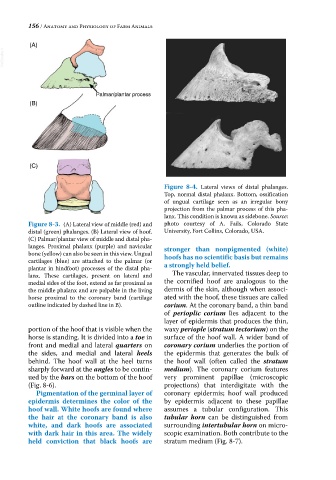
(A)
VetBooks.ir
Palmar/plantar process
(B)
(C)
Figure 8-4. Lateral views of distal phalanges.
Top, normal distal phalanx. Bottom, ossification
of ungual cartilage seen as an irregular bony
projection from the palmar process of this pha-
lanx. This condition is known as sidebone. Source:
Figure 8-3. (A) Lateral view of middle (red) and photo courtesy of A. Fails, Colorado State
distal (green) phalanges. (B) Lateral view of hoof. University, Fort Collins, Colorado, USA.
(C) Palmar/plantar view of middle and distal pha-
langes. Proximal phalanx (purple) and navicular
bone (yellow) can also be seen in this view. Ungual stronger than nonpigmented (white)
cartilages (blue) are attached to the palmar (or hoofs has no scientific basis but remains
plantar in hindfoot) processes of the distal pha- a strongly held belief.
lanx. These cartilages, present on lateral and The vascular, innervated tissues deep to
medial sides of the foot, extend as far proximal as the cornified hoof are analogous to the
the middle phalanx and are palpable in the living dermis of the skin, although when associ-
horse proximal to the coronary band (cartilage ated with the hoof, these tissues are called
outline indicated by dashed line in B). corium. At the coronary band, a thin band
of perioplic corium lies adjacent to the
layer of epidermis that produces the thin,
portion of the hoof that is visible when the waxy periople (stratum tectorium) on the
horse is standing. It is divided into a toe in surface of the hoof wall. A wider band of
front and medial and lateral quarters on coronary corium underlies the portion of
the sides, and medial and lateral heels the epidermis that generates the bulk of
behind. The hoof wall at the heel turns the hoof wall (often called the stratum
sharply forward at the angles to be contin- medium). The coronary corium features
ued by the bars on the bottom of the hoof very prominent papillae (microscopic
(Fig. 8‐6). projections) that interdigitate with the
Pigmentation of the germinal layer of coronary epidermis; hoof wall produced
epidermis determines the color of the by epidermis adjacent to these papillae
hoof wall. White hoofs are found where assumes a tubular configuration. This
the hair at the coronary band is also tubular horn can be distinguished from
white, and dark hoofs are associated surrounding intertubular horn on micro-
with dark hair in this area. The widely scopic examination. Both contribute to the
held conviction that black hoofs are stratum medium (Fig. 8‐7).